Working from home has become the new normal for many Americans during this pandemic. Most are happy without the commute, although working outside the office and in all sorts of strange places, such as bedrooms, backyards, living rooms, has become common place. We’ve had to learn how to work remotely, which in turn means spending a lot more time using computers and smartphones.
Moving out of properly-lit classrooms and offices – and away from ergonomically correct desks – can have long-term effects on our eyes. Excessive time at a computer screen under the best conditions can lead to symptoms of eye strain.
Eye Strain Symptoms include:
- Headaches
- Blurred vision
- Uncomfortable dry eyes
- Neck and shoulder pain
To help avoid eye strain, here are few tips for working from home:
 Record time spent on screens – Most adults age 18 and older spend at least 13 hours each day using digital devices. Extended screen time can cause discomfort and vision difficulties. When working from home, you may lose track of how much time you’re on your computer or smartphone. So keep track of the hours you use devices. That way, you’ll be aware of the demand you’re placing on your eyes.
 Rest and blink your eyes – Researchers found that over 30% of people using digital devices rarely take time to rest their eyes. Just over 10% say they never take a break, even when working from home. The eye muscles get overworked and don’t get a chance to relax and recover. Experts suggest the 20-20-20 rule; every 20 minutes, focus your eyes and attention on something 20 feet away for 20 seconds. You can also get up and walk around for a few minutes.
 Reduce exposure to blue light – In the spectrum of light, blue is more high energy and close to ultraviolet light. So, if you use screens throughout the day, ask your eye doctor about the value of computer glasses that block blue light. Reducing exposure to blue light may help lessen vision problems. At home, using digital devices until bedtime can overstimulate your brain and make it more difficult to fall asleep. Eye doctors recommend no screen time at least one to two hours before going to sleep.
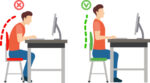 Sit up straight – Proper posture is important. Your back should be straight and your feet on the floor while you work. Elevate your wrists slightly instead of resting them on the keyboard.
- Proper lighting – A setting that’s too bright (sunny backyard) or too dim (cavernous basement) can cause eye strain and headaches. Your screen should be bright enough that you don’t need to squint. A screen shield can help reduce glare.
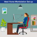 Set up monitor properly – Make sure your computer screen is about 25 inches, or an arm’s length, away from your face. The center of the screen should be about 10-15 degrees below eye level. Cut glare by using a matte screen filter. You can find them for all types of computers, phones, and tablets. Increase font size or set the magnification of the documents you are reading to a comfortable size.
 Consider computer glasses –For the greatest comfort at your computer, you might benefit from having your eye doctor modify your eyeglasses prescription to create customized computer glasses. This is especially true if you normally wear distance contact lenses, which may also become dry and uncomfortable during extended screen time. Computer glasses also are a good choice if you wear bifocals or progressive lenses, because these lenses generally are not optimal for the distance to your computer screen.
- Create Technology-Free Zones – These tips can help reduce eye strain when you’re forced to look at screens all day, creating technology-free zones in certain areas of your home, like the bedroom or bathroom. If you spend the entire day working on the computer, getting in bed and scrolling through social media until you fall asleep won’t do your eyes any favors.
Once you’re done for the day, truly unplug. Read a book or spend some quality time with family members without your phone.
 Get an Eye Exam – If you have tried all these tips and eye strain is still an issue, it might be time to see an eye care professional to schedule an eye exam. The exam may even detect underlying issues before they becomes worse.


 Taking care of your health is critical and you may have concerns related to eye health as a result of the COVID-19 pandemic. The offices of Ophthalmologists and Optometrists are resuming the delivery of comprehensive eye and vision care and implementing new protocols to provide care in a safe and healthy environment.
 Leaving your house these days means a whole new normal. Needing to make sure if you have your mask, your gloves, and hand sanitizer? Now because of masks, millions of eyeglass wearers are discovering the annoyance of mask-induced foggy lenses.
 My daughter Blythe holds binoculars up to her eyes and watches a family of otters play joyously in and out of the kelp. They’re not alone. My wife Patty is laughing as she checks out a group of sea lions in their own game of tag, and the sound of their barking makes me smile. Later that day I’ll hear the appreciative ooh and aah of my family as they watch the sun knife its way into the ocean with its promise to rise again in the morning.