What Is Blue Light?
Blue light, also known as high-energy visible (HEV) light, is a color in the visible light spectrum that can be seen by human eyes. These wavelengths of visible and non-visible light are measured in nanometers (nm), and, in general, the shorter the wavelength, the higher the energy. Blue light is a short wavelength, which means it produces higher amounts of energy.
Unlike other forms of light, the eyes cannot effectively filter blue light, so more can pass through the eye to the retina. Blue light produces both benefits and concerns for our eyes and overall health.
Where Are You Exposed To Blue Light?
The largest source of blue light is sunlight. In addition, there are many other sources:
- Fluorescent light
- Fluorescent light bulbs
- LED light
- Flat screen LED televisions
- Computer monitors, smart phones, and tablet screens
Blue light exposure you receive from screens is small compared to the amount of exposure from the sun. And yet, there is concern over the long-term effects of screen exposure because of the close proximity of the screens and the length of time spent looking at them. According to an NEI-funded study, children’s eyes absorb more blue light than adults from digital device screens.
Benefits of Blue Light?
Moderate amounts of blue light are essential for good health.
- Promotes alertness
- Boosts memory and cognitive function
- Elevates mood
- Regulates circadian rhythm
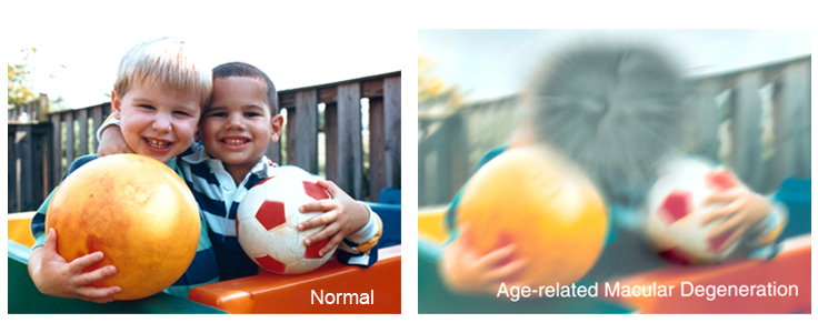
Blue Light and Digital Eyestrain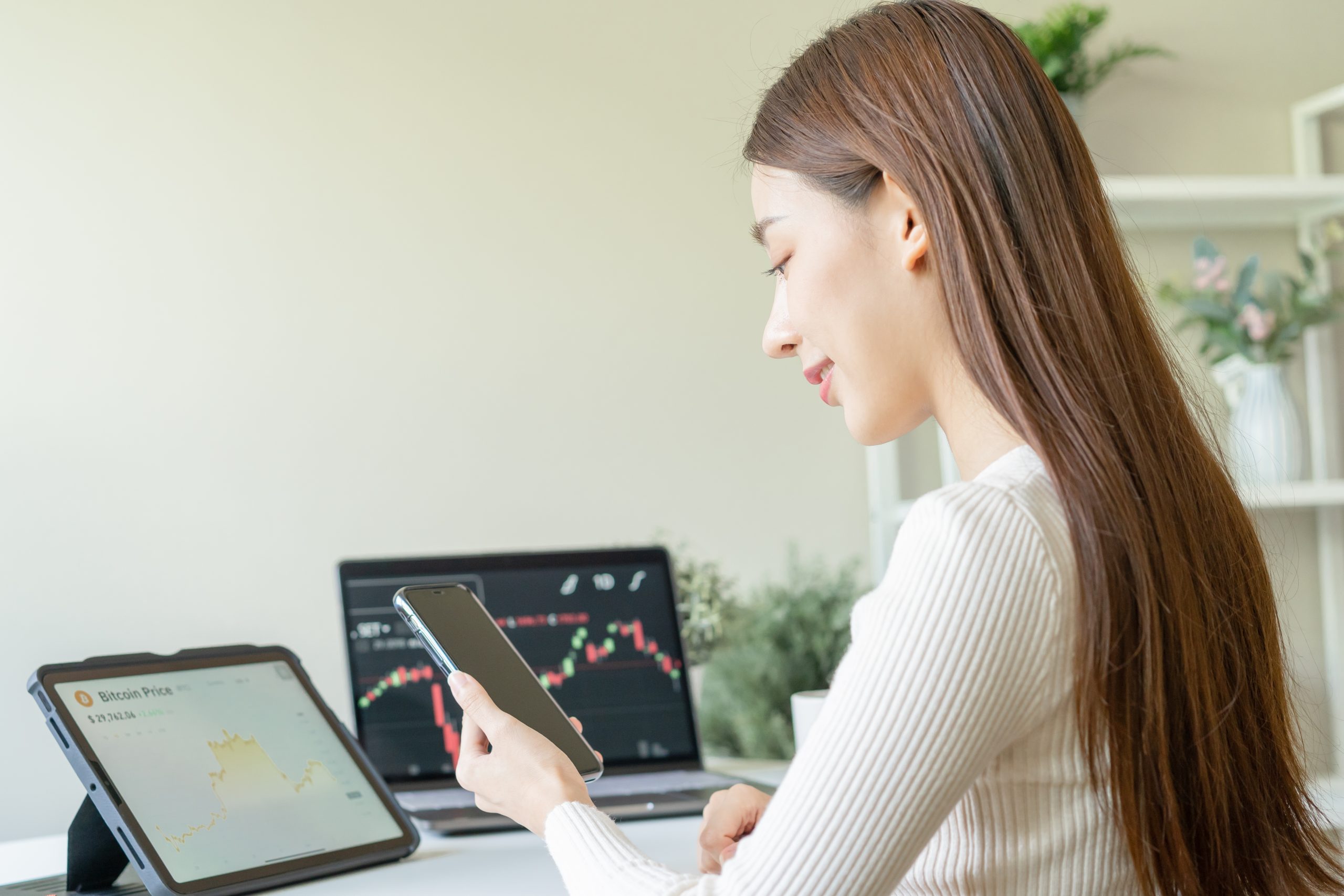
Using digital devices up close or for long periods can lead to digital eyestrain.
Research has shown that when people use computers, laptops, and other digital devices, they tend to blink less often than normal. Fewer blinks can mean less moisture.
Digital eyestrain means different things to different people, but is generally related to the focusing system of the eyes.
When your eyes are strained from staring at a blue-light-emitting screen, you might notice:
- dry eyes
- sore or irritated eyes
- tired eyes
- headaches
- facial muscles fatigued by squinting
Blue light scatters more easily than most other visible light. This may make it difficult for your eye to focus when receiving blue light. Instead, your eye may digest blue light as poorly focused visual static. This reduction in contrast may make it more difficult for your eye to process blue light, potentially contributing to eyestrain.
Still, there isn’t enough evidence to confirm that blue light directly leads to eyestrain. More high-quality studies are needed.
How to Limit Blue Light Exposure
The American Academy of Ophthalmology (AAO) recommends that you take the steps below to reduce digital eyestrain.
1. Practice the 20/20/20 strategy
- While you’re using a device that emits blue light, stop every 20 minutes to focus on objects that are around 20 feet away. Study those objects for 20 seconds before you return to your up-close viewing.
- Keep your eyes moist
- Eye drops, such as artificial tears, and room humidifiers are all good ways to keep your eyes from becoming too dry and irritated while you’re using blue-light-emitting devices.
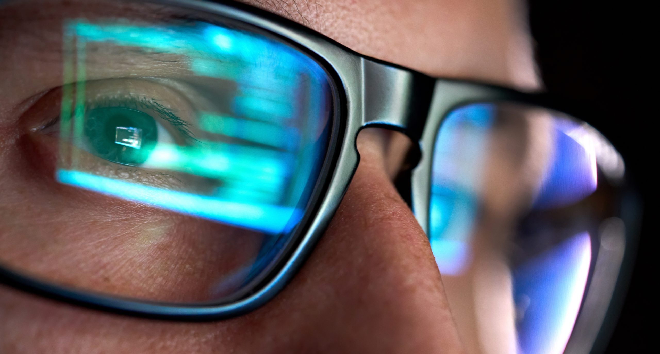
2. Use eyeglasses with the right prescription
Squinting at screens for long periods isn’t recommended for the overall health of your eyes. If you wear prescription eyeglasses to correct your vision, make sure you’re wearing a prescription intended for the distance between your eyes and your screen — ideally an arm’s length away. Most glasses are formulated for longer distances.
3. Adjust the blue light on your screen
To lower the risk of eyestrain and sleep disturbance, you may want to set your screens to a “night shift” setting with warmer tones. You can also purchase blue-light-filtering screens to slip over your computer screen when you’re working at night. The filter could cut the glare of your screen.
And a 2020 research study shows that they block 30 to 60 percent of blue light, though it isn’t clear whether blocking the blue will help preserve the sleep-wake cycle for those who use backlit screens before bedtime.
 Harsh weather conditions can reduce the natural moisture in your eyes and the irritation usually results in a burning or itching sensation that often leads to rubbing or scratching your eyes which can worsen the symptoms. Sometimes it feels like there is a foreign object in your eye and for some, dry eyes can even cause excessive tearing, as your eyes try to overcompensate for their lack of protective tears. Prolonged, untreated dry eyes can lead to blurred vision as well. Between the harsh winter winds outside and the dry heat radiating inside, our eyes are very quickly irritated and dried in the winter months. The result is itchy, dry eyes that may cause pain, blurred vision, a burning sensation, or even watery vision as our eyes try to compensate for the dryness.
 Make sure they take frequent screen breaks. Instead of focusing directly on the screen, encourage your child to look around the room every now and then, or take some time to stare out the window (at least 20 seconds is recommended by the American Optometric Association). You can even remind them to blink.
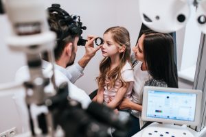 It’s easy for us to forget about our eyes let alone our child’s, but it is very important to get your child’s eyes checked regularly.
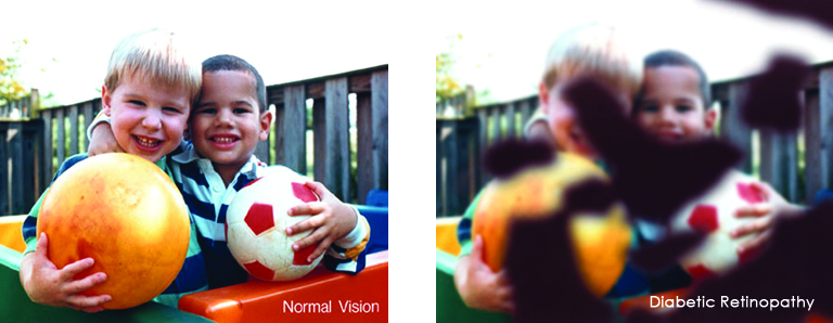
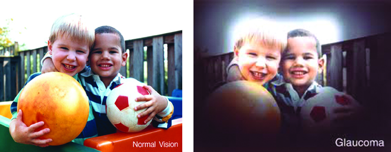
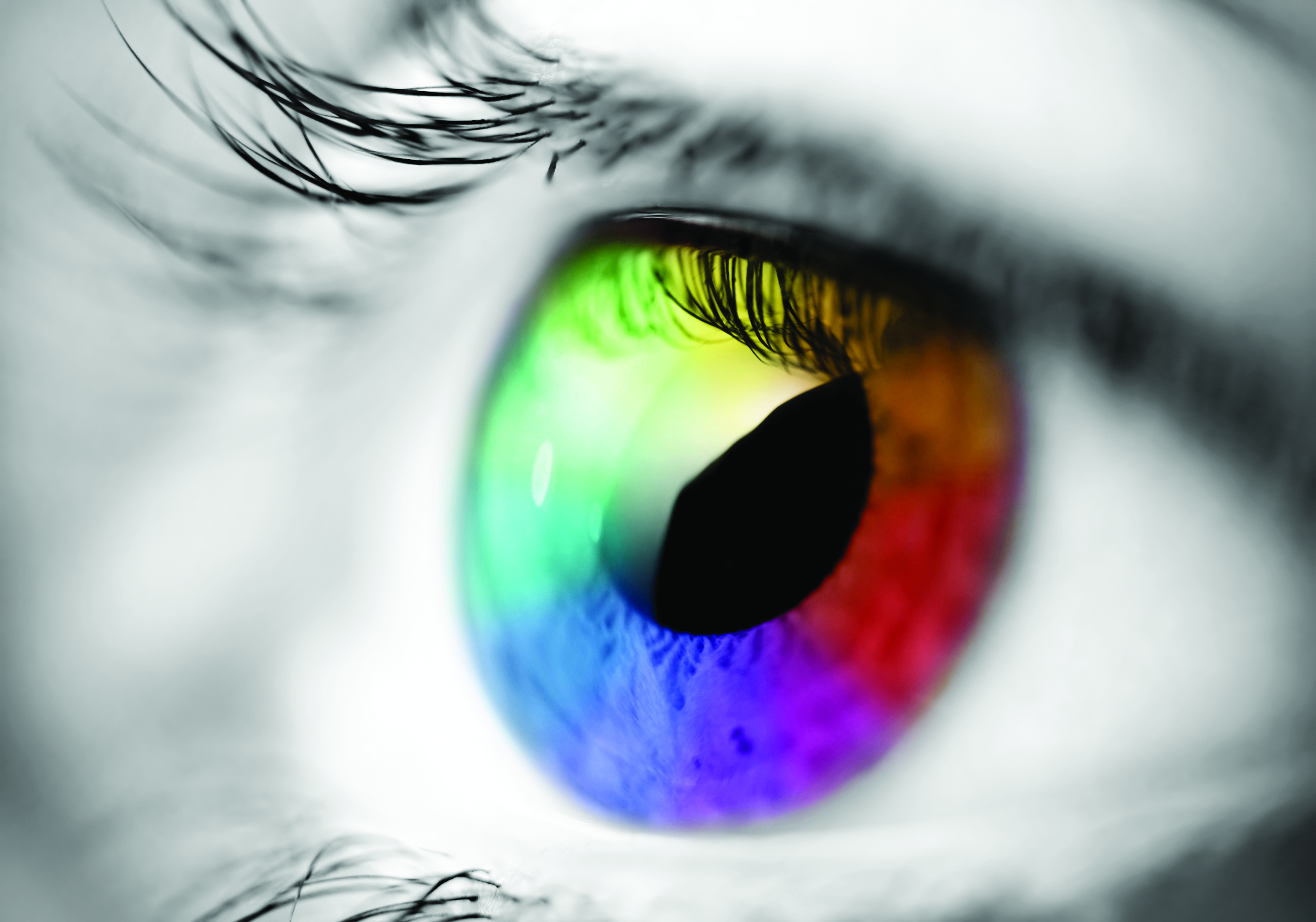 Most people have eye problems at one time or another. Some are minor and will go away on their own, or are easy to treat at home. Others need a specialist’s care. Some eye issues come with age while others may be a serious condition.
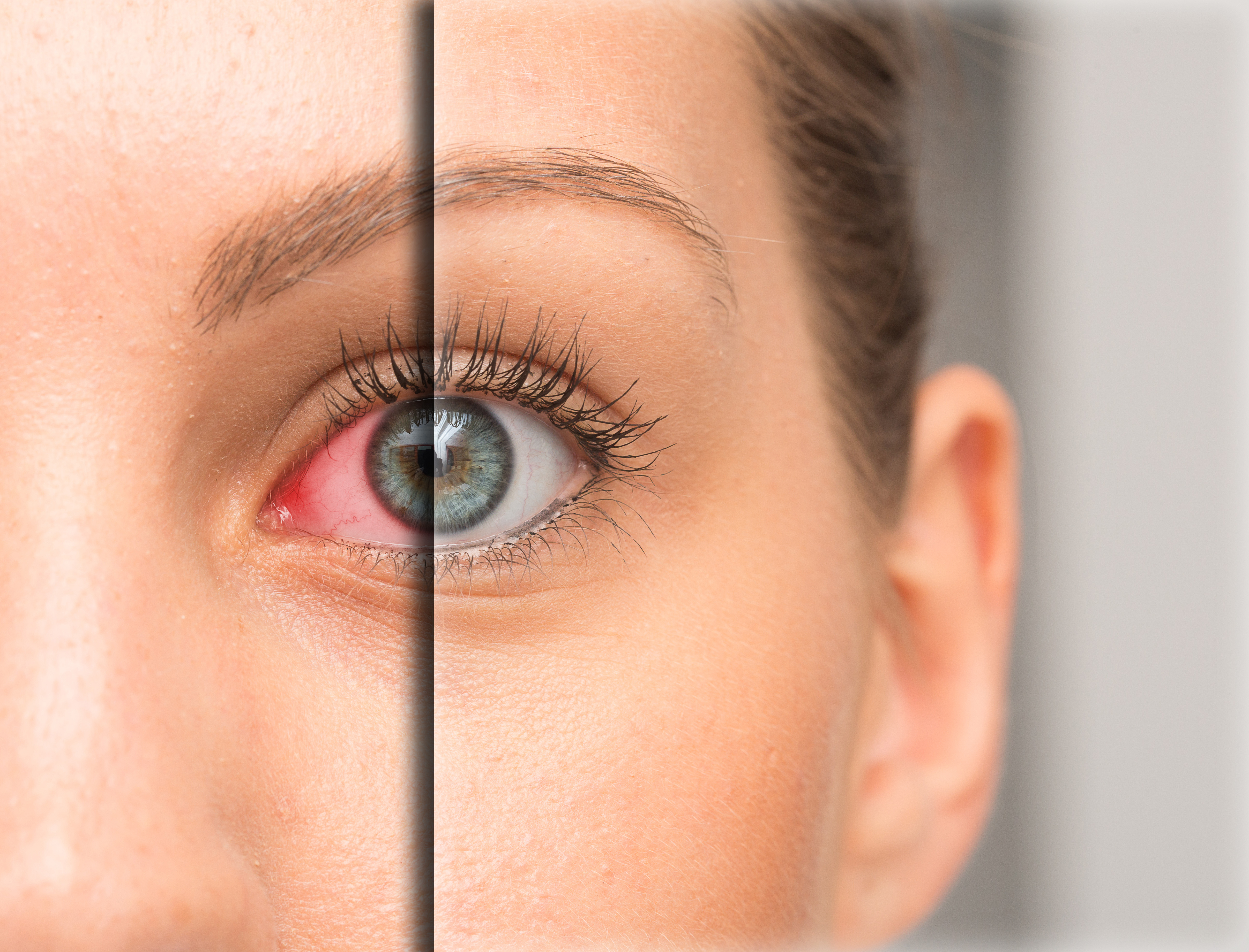 Dry eye is a common condition that occurs when your tears aren’t able to provide adequate lubrication for your eyes. Tears can be inadequate for many reasons. For example, dry eyes may occur if you don’t produce enough tears or if you produce poor-quality tears. Dry eyes can also feel very uncomfortable.
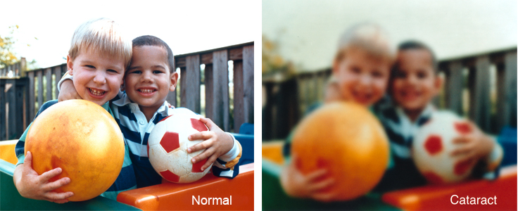
 As winter shifts to spring, and flowers, grasses and trees begin to bloom, spring can take a toll on your eyes if you suffer from seasonal allergies. The spring season has a marked an increase in pollen and allergens in the air, that leave you with congestion, headaches, and itchy, swollen eyes, known as eye allergies.