What To Expect On Your Next Visit To Your Eye Care Practitioner During COVID-19 Pandemic

Taking care of your health is critical and you may have concerns related to eye health as a result of the COVID-19 pandemic. The offices of Ophthalmologists and Optometrists are resuming the delivery of comprehensive eye and vision care and implementing new protocols to provide care in a safe and healthy environment.

While changes vary from state-to-state as well as individual practice locations, patients should expect that their eye doctors, like all medical professionals, are adhering to federal, state and local health directives regarding infection prevention measures and implementing appropriate safety procedures to reduce the risk of COVID-19 transmission within the office. These not only include strict protocols for cleaning and sterilization, but measures to effectively manage patient flow and encourage physical distancing. Patients should expect screenings for symptoms of COVID-19 and taking patients’ temperature upon their arrival at the facility, limiting the number of guests allowed in waiting rooms and requiring everyone to wear a facemask and/or gloves before entering the office.

Prior to the appointment, you should be asked a series of questions such as whether you have been diagnosed with COVID-19, whether you have been exposed to anyone who has an active COVID infection, and whether you have any of the typical symptoms of a COVID-19 infection including fever, cough, shortness of breath, or some impairment of your sense of taste or smell. You may also be asked about recent travel history. If there is a concern that you may have COVID-19 infection, then the appointment may be rescheduled to a later date.

Once you get to the office, you will be interviewed again to see if you have any symptoms suggestive of COVID-19 infection. You may be asked to have your temperature taken with a non-contact thermometer and to wash your hands or scrub your hands with hand sanitizer before entering the office. You may be asked not to bring any family members or assistants with you.

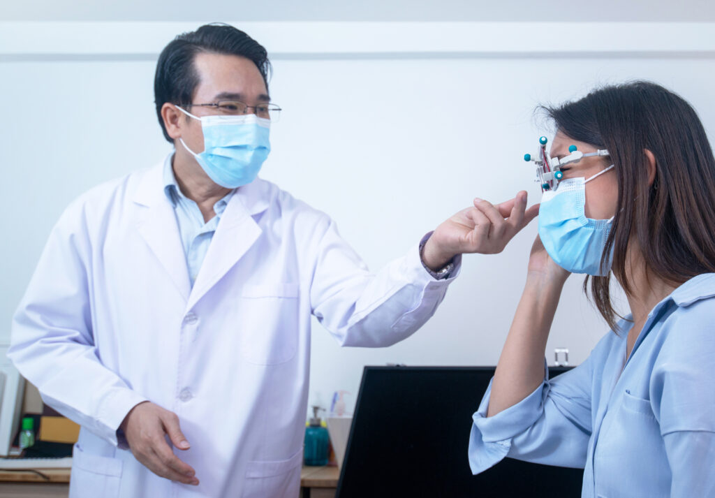
Every patient (and anyone accompanying the patient) is required to wear a mask, and the mask must be worn properly, covering both the nose and mouth. The mask must be worn through the entire visit, and conversations with the doctor or staff will be limited. The tests that are used to monitor glaucoma may be performed with modified protocols to minimize the time you spend at the office.

How to Prepare for Your Eye Doctor Visit

  • Don’t go if you’re feeling sick. If you wake up with a cough or a sore throat, it’s probably best to reschedule your appointment and call your primary physician. Your routine eye exam can wait. Don’t be surprised if someone takes your temperature upon arrival to ensure safety for everyone.
  •  Fill out forms in advance. If possible, fill out your intake forms online before your appointment. This will limit your face-to-face contact and your overall time within the office. Any additional information that’s requested by your doctor can also be done over the phone.
  • Follow social distancing guidelines. There may be new procedures in place to help with social distancing, from markers on the ground indicating where to stand, to a limited number of people allowed in certain areas. Depending upon the office location, you may need to call from your car to check in. Every office is different, so be sure to check with your doctor’s office about their requirements.
  • Go alone to your appointment. Most offices are only allowing the patient to enter the office, with the exception of those who require a caregiver or a guardian to attend the appointment with them. This limits the amount of people going in and out of the office, and helps maintain a more sterile, clean environment.
  • Wear a mask to your appointment. Depending on your state, a mask may either be mandated or highly recommended when you are in public places. The Centers for Disease Control and Prevention highly advises people to wear a mask covering their nose and their mouth in order to help stop the spread of COVID-19.

As your eye care doctors begin reopening and operations proceed under a “new normal,” your ophthalmologist and/or optometrist are working to ensure the continued safe delivery of essential eye care during the COVID-19 pandemic.

To know exactly what to expect during your appointment, call your doctor’s office to find out how to best prepare for your visit.

Beauty is in the Eyes of the Beholder

I love the idea that beauty is in the eyes of the beholder. It reminds us that individuals can make choices about what they perceive to be true beauty.

Let’s consider art… one person’s beautiful can be another person’s junk. If beauty is in the eyes of the beholder, the question is – are we taking the time to appreciate all that surrounds us? From Mother Nature’s unlimited panoplies of possibilities to the innocent smile of a child, beauty is the catalyst that makes being alive the greatest of all human adventures.

I am sure you’ve heard the phrase “stop and smell the roses,” so why not take the time to stop and observe the beauty your eyes can behold.

The Discovery Eye Foundation is working every day to preserve your vision and give all of us the chance to see what’s truly beautiful and treasure it.  All you have to do is open your eyes and take a look.

Louis Armstrong said it well:

I see trees of green and red roses too.
I see them bloom for me and you.
And I think to myself,
what a wonderful world.

You’re right Louis… it is a wonderful world if we all just keep appreciating the beauty that’s out there for all of us.

 
Donate today to help support the Discovery Eye Foundation! 

DonateNow

Tom Sullivan
DEF’s Ambassador of Vision

sullivanvision.com


June is Cataract Awareness Month

June is Cataract Awareness Month. This is a time to raise cataract awareness and help to educate people about one of the leading causes of treatable vision loss in the United States. There are 24 million Americans over the age of 40 who are affected by cataracts, so it seems fitting that an entire month should be dedicated to cataract education and awareness.

A cataract is a clouding of the eye’s lens, which blocks or changes the passage of light into the eye. The lens of the eye is located behind the pupil and the colored iris, and is normally transparent. Vision may become blurry or dim because the cataract stops light from properly passing through to the retina. Generally, a cataract does not cause pain, redness or tearing, but does cause increasing difficulty in seeing clearly.


Here is an overview of Cataracts:

View Video


Cataract symptoms:

  • Cloudy
  • Night vision
  • Glare
  • Halo
  • New glasses
  • Yellow tint
  • Double vision

Some Risk factors for cataracts include:

  • Older age
  • Intense heat or long-term exposure to UV rays from the sun
  • Certain diseases, such as diabetes
  • Inflammation in the eye
  • Hereditary influences
  • Long-term steroid use
  • Eye injuries
  • Eye diseases
  • Smoking

Cataracts can be easily diagnosed and visiting your eye doctor regularly is important in helping protect your eyes from further damage. An annual eye exam is recommended for everyone over the age of 60, and bi-annual exams for adults between 41-60 years to check for developing eye or vision problems.


There are a few treatment options, below is one of the newest options:
View Video


There is no proven way to prevent age-related cataracts. However, choosing a healthy lifestyle can slow the progression of cataracts. Some ways to delay the progression of cataracts include avoiding smoking, reducing exposure to UV rays, eating healthy foods, and wearing proper eye protection to avoid eye injury.

For more information and where you can find support for Cataracts click here .

Coronavirus and Your Eyes: What you should know

Coronavirus (COVID-19) can cause mild to severe respiratory illness. Symptoms such as fever, cough, shortness of breath and pink eye can show up 2 to 14 days after a person is exposed. People with severe infections can develop pneumonia and even die from complications of the illness.

To cut your personal risk of contracting the COVID-19, avoid touching your eyes, nose, mouth and face with unwashed hands. It is the mucous membranes (membranes that line various cavities in the body) that are most susceptible to transmission of the virus.

To avoid infecting others with the coronavirus, the Centers for Disease Control and Prevention (CDC) has recommended the use of face masks to be worn when out in public. Face masks can reduce the spread of coronavirus by people who are infected but have no symptoms of the virus (asymptomatic). Face masks, however, do not protect your eyes from infection.

Here are a few tips on how to protect yourself and others:

1. Practice safe hygiene and social distancing — The Centers for Disease Control and Prevention (CDC) offer these general guidelines to slow the spread of disease:

  • Wash your hands often with soap and hot water for at least 20 seconds. (Singing Happy Birthday twice is about 20 seconds)
  •  You should especially wash your hands before eating, after using the restroom, sneezing, coughing or blowing your nose.
  • If you can’t get to a sink, use a hand sanitizer that has at least 60% alcohol.
  • Avoid touching your face — particularly your eyes, nose, and mouth.
  • If you cough or sneeze, cover your face with your elbow or a tissue. If you use a tissue, throw it away promptly. Then go wash your hands.
  • Avoid close contact with sick people. If you think someone has a respiratory infection, it’s safest to stay 6 feet away.
  • Stay home when you are sick.
  • Regularly disinfect commonly touched surfaces and items in your house, such as doorknobs, refrigerator door handles and counter tops.

2. Coronavirus may cause pink eye, so avoid touching eye discharge — Someone may have pink eye but it doesn’t mean that person is infected with coronavirus. But a recent study suggests that up to one third of people hospitalized with coronavirus experience eye problems, such as viral pink eye or conjunctivitis. It’s important to know that the virus can spread by touching fluid from an infected person’s eyes, or from objects that carry the fluid.

3. Avoid rubbing your eyes — We all tend to do it and natural habits can be hard to break but doing so will lower your risk of infection. Use a tissue instead of your fingers when you feel the urge to rub your eye or even to adjust your glasses. Dry eyes can lead to more rubbing, so consider adding moisturizing drops to your eye routine. If you must touch your eyes for any reason wash your hands first with soap and water for at least 20 seconds. Then wash them again afterwards.

4. If you wear contact lenses, consider switching to glasses for a while — There’s no evidence that wearing contact lenses increases your risk of coronavirus infection although contact lens wearers touch their eyes more than the average person. Substituting glasses for contacts can decrease irritation and will lower the chances of you touching your eye. If you choose to continue wearing contact lenses, follow these hygiene tips.

5. Make sure you are well supplied on eye medicine prescriptions if you can — During this pandemic experts have advised patients to stock up on critical medications, enough to get by if you are quarantined or if supplies become limited. If your insurance allows you to get more than 1 month of essential eye medicine, such as glaucoma drops, you should do so. Some insurers will approve a 3-month supply of medication in times of natural disaster. Ask your pharmacist or ophthalmologist for help if you have trouble getting approval from your insurance company. Don’t wait until the last minute to contact your pharmacy, request a refill as soon as you’re due.


IF YOU’RE NOT FEELING WELL – Call your family doctor. If you suspect you may have pink eye (conjunctivitis), call an eye doctor near you. It is suggested that patients not go directly to medical or eye care facilities without a prior phone call to help to decrease the possible spread of the virus. A phone call allows the health facility to prepare for your visit and diagnose and treat you in a proper manner.
You may feel nervous about going to the doctor’s office during this pandemic. But treatment for eye emergencies should not be delayed. Ophthalmologists, like all medical professionals, follow strict hygiene and disinfection guidelines.

Ophthalmologists are available to treat urgent eye issues, deliver eye injections and provide critical care. Call your ophthalmologist or other medical doctor as soon as possible in the following situations:

  • You have macular degeneration or diabetic retinopathy and get regular eye injections.
  • You notice changes in your vision (like blurry, wavy or blank spots in your field of vision).
  • You experience an eye injury, even if it seems minor.
  • You suddenly lose some vision.
  • You have eye pain, headache, red eye, nausea and vomiting.

Protect yourself and the eye care team by following these precautions:

  • Wear a mask to a medical appointment. The mask should cover your nose and mouth.
  • If you have a cough or a fever, or have been in close contact with someone who has these symptoms, you must call your doctor’s office ahead of time and let them know. After you speak with the health care profession, it may be decided that your visit is not an emergency, and you can be treated at home. If you arrive sick at the doctor’s office, you should wear a protective covering or mask, and they may want you to wait in a special room away from other patients.
  • If you need to cough or sneeze during your exam, move back from the microscope. Bury your face in the crook of your arm or cover your face with a tissue. Wash your hands with soap and water right away.

For more Coronavirus information visit: www.cdc.gov/coronavirus

For more information about the Discovery Eye Foundations new research for Coronavirus vaccine visit: www.discoveryeye.org/covid-19-emergency-research/

COVID-19 EMERGENCY RESEARCH

DonateNow

A MESSAGE FROM ANTHONY B. NESBURN, DEF PRESIDENT & MEDICAL DIRECTOR


The world is being held hostage by coronavirus (COVID-19). The number of cases and deaths are climbing everyday. Scientists and pharmaceutical companies are working feverishly to create a vaccine, but it is at least a year away. There is also hope that existing FDA-approved drugs, such as hydroxychloroquine and azithromycin, may be able to ameliorate the disease and prevent deaths.  These need to be tested now—see below.

During this emergency, DEF Research Director Dr. Cristina Kenney’s laboratory is applying the knowledge gained from her work on age-related macular degeneration to join the search for drug(s) that may be an immediate bridge to treat COVID-19.

Much of Kenney’s research focuses on mitochondria, which play a vital role in disease and death from infections such as COVID-19. Mitochondria may help explain the severity of disease and response to treatment in different ethnic populations and age groups.

Kenney will focus on two crucial COVID-19 studies using her unique laboratory systems:

  1. Determine which promising experimental treatments for COVID-19 patients are most likely to benefit which patients.
  2. Determine the role of mitochondria in susceptibility to severe COVID-19 disease and death in different ethnic/racial populations and age groups.

UPDATE: DEF Researchers Making Progress in the Fight to Defeat COVID-19


Join us and others in supporting this work. At this critical juncture, any financial help you are able to provide to our vital research efforts to stop the scourge of coronavirus is greatly needed and deeply appreciated. 

Click here to  DONATE BY MAIL

Click to DONATE ONLINE:DonateNow


For more information on COVID-19 go to www.coronavirus.gov

OUR EYES WORK LIKE CAMERA’S!

The inner workings of the human eye are complex, but at the same time, fascinating. Have you wondered how exactly they do work or what are the major parts of the eyeball involved in creating vision? Let’s find out.

These tiny cameras spend every day processing millions of pieces of information at lightning fast speeds, and turn them into the simple images we see almost instantly.

In reality, this process is anything but simple. The eye has several distinct parts, each of which has specific responsibilities that work together like a machine.

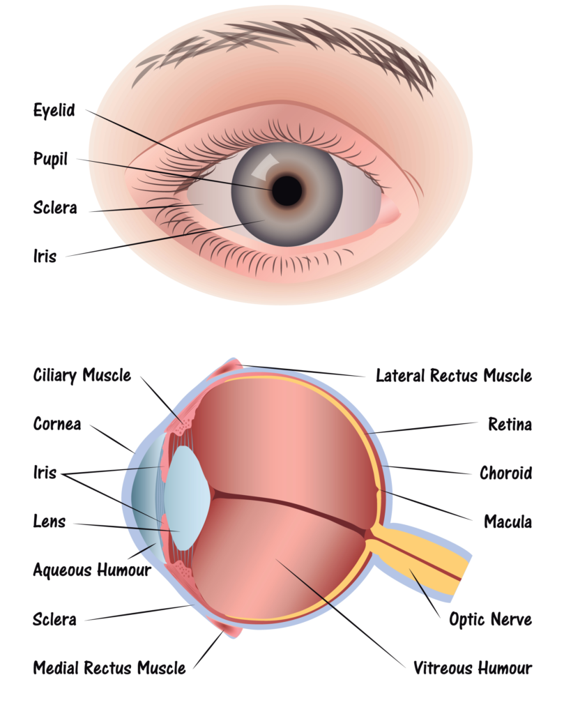
The eyeball is just like a camera. In fact, human eyes are part of a classification known as “camera-type eyes.” And just like a camera, it can’t function without the presence of light.  As light hits the eyes, it’s focused by the eye in a way similar to a camera lens. This process allows the images we see to appear clear and sharp rather than blurry.

There are specific parts of the eye that make this focusing process possible. Each beam of light that hits the eye goes through a series of steps:

Step 1: Light passes through a thin layer of moisture
Step 2: Light hits the cornea.  The cornea is transparent, and is the first layer to begin focusing light within the eye. The cornea is connected to the sclera, which is a tough fiber on the outside of the eye that acts as protection.

Step 3: Behind the cornea is another liquid layer known as the aqueous humor, and its job is to maintain pressure levels in the front of the eye as light is passing through.

Step 4: Once light has passed through the aqueous humor, it has finally reached the pupil. The pupil is the round entryway of the colored iris.

Step 5: Once the pupil determines how much light it will let inside your eye, the job passes to the lens. The lens factors in the amount of light the pupil lets in, and figures out how far away you are from the object that the light is reflecting off of, or the object you’re trying to see. From there, the lens focuses your image into an accurate view of what you’re looking at. Part of this process is controlled by muscles in the lens called ciliary muscles, which expand and contract to pull on the lens and allow it to focus properly.

Step 6: As light reaches the center of the eye passes through another layer of moisture, called the vitreous, or vitreous humor. Then, it reaches the final stop in the process: The Retina.

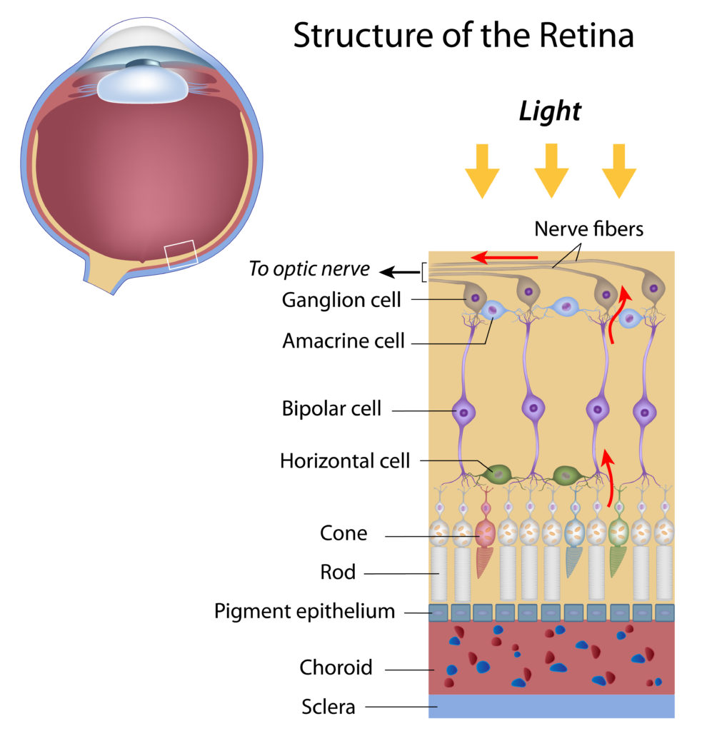
The retina is the back of the eye. If the lens in your eye is most like a camera, the retina is most like its film – this is where the final product is projected. The retina has several parts:

  • Macula: The center of the retina. The center point of the macula is called the fovea, and it has the most photoreceptors and nerve endings of any part of the eye.
  • Photoreceptors: Split into two designations – rods and cones.
    • Cones are in the macula. When there is bright light, cones provide clear, sharp central vision and detect colors and fine details.
    • Rods are located outside the macula and extend all the way to the outer edge of the retina. They provide peripheral or side vision. Rods also allow the eyes to detect motion and help us see in dim light and at night.

  • Retinal pigment epithelium: Abbreviated RPE, this is a tissue layer below the rods and cones which absorbs any extra, unneeded light.
  • Choroid: The choroid is behind the retina, and is in charge of making sure the retina and RPE have enough nutrition flowing from small blood vessels.

Once the photoreceptors have converted light into an electronic signal, they send a signal to the brain’s visual command center and you have vision. It’s amazing what even small parts of our bodies can do.


How the Eye Works

January is Glaucoma Awareness Month

National Glaucoma Awareness Month reminds all of us to get regular eye exams and show support for those suffering from this conditionGlaucoma is one of the leading causes of vision loss and blindness.  Glaucoma is often called “the sneak thief of sight” because glaucoma has few symptoms or warning signs in its early stages. It can be diagnosed only by a full eye exam by an eye care practitioner.

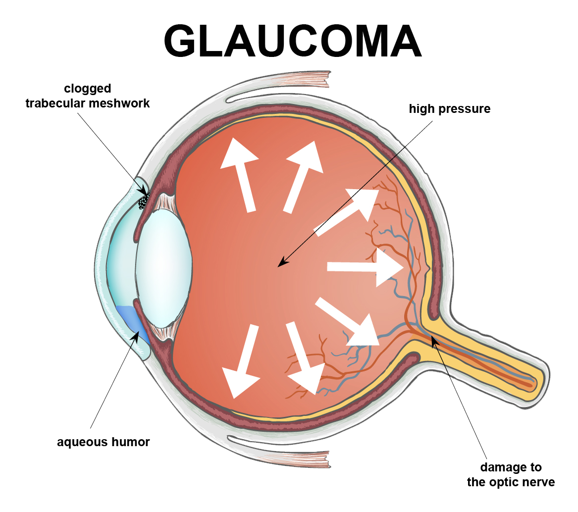
Glaucoma is a disease where pressure builds up and damages the eye’s optic nerve. Types of this disease include the common Primary Open Angle Glaucoma, which causes peripheral eyesight to slowly diminish and is age-related. Angle Closure Glaucoma, where the fluid drainage system is narrow and closed so that the aqueous fluid remains in the front chamber of the eye and intraocular pressure rises; and Low Tension Glaucoma, where the optic nerve becomes damaged in spite of the intraocular pressures being within the normal range. There’s currently no way to restore vision lost from glaucoma because once the nerve cells become damaged, they do not regenerate.


A few important facts you should know about Glaucoma in adults:

  • More patients than ever are affected – Over 3 million people in the U.S. have glaucoma, and the number is rising.
  • Glaucoma can affect people of all ages – The most common form of glaucoma, Primary Open Angle Glaucoma, becomes more prevalent with increasing age. However, glaucoma can strike anyone, even infants and children but it is rare.
  • Demographics do play a role – Glaucoma is a leading cause of blindness among African Americans; it’s also highly prevalent in Hispanics over the age 65.
  • Is glaucoma hereditary? The risk of developing Primary Open–Angle Glaucoma is up to nine times more likely if parents or siblings have the disease.
  • Hope for future glaucoma patients – Although there is no cure for any form of glaucoma, early diagnosis and treatment help control the disease and slow the process of vision loss or blindness.

Newer Glaucoma Treatments 

Glaucoma treatment usually begins with the use of topical (eye drop) medications which lower the intraocular pressure. Within the past two years, two new topical medications have been approved for the treatment of glaucoma: VYZULTA® and Rhopressa®. VYZULTA® is a modification of a class of medications currently used to treat glaucoma – the prostaglandin analogs. This drug helps lower intraocular pressure by increasing the drainage of fluid (aqueous humor) from the eye.

Rhopressa® is part of a new class of drugs used to treat glaucoma called Rho kinase inhibitors. Rhopressa®, like VYZULTA®, also lowers intraocular pressure by increasing aqueous outflow. Both Rhopressa® and VYZULTA® are dosed once daily and pose few, if any, systemic safety concerns.
 

Laser

Using a laser to make a small opening in the iris to help with fluid drainage usually cures Angle-Closure Glaucoma.  This procedure is called a laser peripheral iridotomy.

For Primary Open Angel Glaucoma, when eye drops are not enough to reduce the pressure then a procedure called Selective Laser Trabeculoplasty (SLT) can be used. The SLT reduces intraocular pressure by stimulating increased outflow of fluid from the eye. SLT offers an improved safety profile compared to older glaucoma laser therapies and may lower eye pressure by as much as 20 to 30 percent. It is typically used as the next step in patients whose glaucoma is uncontrolled on medical therapy. Because of its excellent benefit-to-risk profile, however, SLT can sometimes be used in place of medications, especially in patients who have difficulty with their eye drops.
 

Surgery

When other treatments fail, there are many surgical therapies to lower the eye pressure. These surgical approaches, which are riskier than medical therapy or lasers, are usually employed when non-surgical means do not work well enough to stop vision loss.
 

MIGS

The goal of Minimally Invasive Glaucoma Surgery (MIGS) is to reduce intraocular pressure by enhancing the eye’s own internal aqueous humor drainage system. Some MIGS can be performed as stand-alone procedures, while others are typically done along with cataract surgery in patients with visually significant cataracts and mild to moderate glaucoma.

There are now a variety of recently FDA approved MIGS available for use in this country. MIGS typically are performed through a small incision in the eye with minimal tissue trauma and offer a favorable safety profile as well as more rapid visual recovery than traditional glaucoma surgery. There are many well qualified glaucoma specialists that perform the MIGS procedure.


Glaucoma Treatment Overview


National Glaucoma Awareness Month reminds all of us to get regular eye exams. Don’t let glaucoma steal your sight!  The best way to protect your sight is to get an annual comprehensive eye examination.


Thanks to funding from private philanthropists, DEF’s research continues to make great strides toward cures and treatment for glaucoma.  If you would like to support DEF’s sight saving research please donate today!

 DonateNow

A Vision of Christmas

A special holiday message from our beloved blind Ambassador of VisionTom Sullivan

My wife Patty loves every aspect of the Christmas season, and she begins celebrating the yule tide even before Thanksgiving.  The transforming of our house with traditional decorations brings her a marvelous sense of family and holiday season.  She loves to look at every ornament remembering how we acquired it.  As the lights turn on outside our house, she is busy making sure that every bulb on our tree is placed just so to give the room a warmth that in every way reflects her commitment to the true spirit of the season.

While I am listening to football upstairs, she’s singing along with Celine Dion as she completes the visual picture that for Patty confirms that there really are important traditions that make our home a place of love, beauty, and peace.  For the record, let me say that I really do enjoy Christmas even though I offer half-hearted complaints that Patty plays carols starting even before Turkey Day.  The truth is…I love them just as much as she does.

For over thirty years we went to Colorado every Christmas to ski, and those experiences were wonderful to just be in the mountains and feel the cold nipping at your nose as you fly down a ski slope.  We often would take a sleigh ride on Christmas Eve with hot chocolate spiced with peppermint schnapps.  Listening to a children’s choir and being with people we love always seems to make things a little more right with the world.  So I really do love Christmas, but when I consider Patty’s ebullient joy in the visuals of the holiday, I admit it makes me just a little sad to know that I’ll never be able to see them.

I realize that people go to extraordinary lengths in their decorating zeal and that certainly some of their elaborate choices that brighten the neighborhood could be considered over the top, but when Patty’s carols are playing and she’s enjoying the happiness of singing along with the decorations just so, I know that what she’s reflecting is her true commitment to her faith and the celebration of the season that she treasures. 

So open your hearts and your eyes and take in the true experience of the Christmas spirit.  It’s my hope that you’ll consider supporting Discovery Eye Foundation’s commitment to preserving vision allowing more people every year to enjoy not just the smell of a Christmas tree, or the taste of a Christmas toast, the touch of a hug from a loved one on Christmas morning, or the beautiful sounds of Silent Night sung by the voice of a child.

From all of us at Discovery Eye, we wish you a Merry Christmas and Happy Holidays!


Tom Sullivan
DEF’s Ambassador of Vision

sullivanvision.com

Common Eye Problems in Winter

Winter time is here!! And as most people would expect, the holidays are a very popular time of the year. But the winter also comes with extreme temperatures, humidity and precipitation, so don’t let it affect your vision!

Be on the lookout for these common eye conditions this winter and hopefully, you will comfortably enjoy the season without any problems.


DRYNESS

Cold outdoor air and heated indoor air often have less moisture than other seasonal environments. In the winter, you may experience dry skin, chapped lips, and dry eyes due to this low humidity. Cold winter winds may also dry your eyes out. To learn more about dry eyes, visit the linked website at Dry Eyes.

To minimize the drying ability of winter air, keep yourself hydrated. Use non-preserved artificial tears several times a day. Running an humidifier in your home to improve the quality of your indoor air will help as well.


EXCESS TEARING

While some people experience a lack of tears in the winter, others have the opposite problem. Excess tearing and runny eyes can occur due to cold air, biting winds, or seasonal allergies. Pay attention to when your eyes tear up to determine the cause. If your eyes start to water when you step outside or when the wind blows your way, wear sunglasses or goggles to protect your eyes. Although it seems strange using additional artificial tears for teary eyes, nonetheless they can lessen this reflex tearing.

If you experience excess tearing and itching while indoors, try an allergy medication and appropriate eye drops to reduce the effect of seasonal allergies. If you cannot determine the cause of your watery eyes or if over-the-counter treatments have no effect, especially if the wateriness alters your vision, see an eye doctor.


LIGHT SENSITIVITY

Winter skies can seem dark and gloomy, but snowfall and ice create many reflective surfaces that can dramatically increase the amount of light that reaches your eyes. If you have sensitive eyes, you may experience even more blinking, discomfort, tearing and other symptoms in bright winter light.

Some individuals develop new light sensitivity during winter due to a condition known as “snow blindness”.  Always protect your eyes with UV filtering glasses or ski goggles when going outdoors for long periods of time, including when walking, shoveling snow, or other winter activities.


REDNESS

Harsh winter conditions can cause redness, tenderness, and inflammation in the eye area. You may have swollen eyelids or redness over the normally-white part of the eye (the conjunctiva which covers the white sclera).

This redness could result from dry eye or seasonal allergies. Use non-preserved artificial tears every 2-3 hours.  To reduce the discomfort of inflamed eyes, apply a cool compress, such as a damp washcloth and take an over-the-counter oral pain medication.  If your symptoms persist, see an eye doctor to determine the cause of the irritation.


VISION CHANGES

While many winter eye health problems result from increased light or decreased moisture, you can also experience eye conditions caused by cold temperatures.

If you notice vision changes while out in the cold, move to a warm area as soon as possible. Use non-preserved artificial tears every 2-3 hours to to keep your eyes moist. If your normal vision doesn’t return after 30 minutes or so, seek medical attention.


If you experience any of the seasonal problems listed above for a prolonged period, consult your eye doctor.

Your EYES will be thankful for Thanksgiving Dinner!

Thanksgiving is almost here; a meal that nourishes the family bonds and traditions.  It’s the one time of the year where you can guarantee your eyes will be bigger than your stomach. This meal also has another added bonus — almost every item on the Thanksgiving table is healthy for your eyesight!

Here are several of the most popular Thanksgiving dishes and their corresponding benefits to your eye health:


Turkey –

No Thanksgiving is complete without a turkey, roasted golden brown and stuffed with fresh vegetables and herbs. Turkey is loaded with zinc and B-vitamin niacin, which helps prevent the formation of cataracts. Cataracts are the leading cause of vision loss in the United States, so gobble up!  You are “preventing” cataracts with every bite.


Spinach, Green Bean Casserole, Asparagus and Brussel Sprouts –

These foods and other leafy greens are loaded with lutein and zeaxanthin, two nutrients that protect the retina, which may also help reduce the risk of cataracts and macular degenerationHealthy Green Bean Casserole Recipe


Sweet Potatoes –

Sweet potatoes are full of Beta-carotene, which is a carotenoid and antioxidant that promotes night vision and overall good eyesight. Sweet potatoes are also loaded with vitamins C and E. Diets that are rich in these vitamins can help prevent or delay the development of cataracts and macular degeneration. Mashed Sweet Potatoes Recipe


Cranberry Sauce –

Cranberries contain bioflavonoids, a large class of antioxidants. Bioflavonoids are found in the pulp, skin and rinds of foods that contain vitamin C. Both flavonoids and vitamin C help protect the eyes from free radical damage caused by pollution and the body’s normal metabolic processes.


Pumpkin Pie –

Pumpkin is one of the best sources of vitamin A, so pumpkin pie is an eye-healthy dessert. In fact, one cup of cooked, mashed pumpkin contains more than 200 percent of the recommended daily intake of Vitamin A. Vitamin A provides nourishment and protection to the eye’s lens, cornea and macula (part of the retina), so it improves night vision. 


A diet that’s full of the right nutrients is a great start to keeping your eyes healthy, but don’t forget that regular eye exams are equally important! We hope you enjoy a happy and delicious Thanksgiving with family and friends.

*For more eye healthy recipes click here EYE COOK