The Brain and the Eye – How They Work Together

The Brain and the Eye

The eye works like a camera. The iris and the pupil control how much light to let into the back of the eye, much like the shutter of a camera. When it is very dark, our pupils get bigger, letting in more light; when it is very bright our irises constrict, letting in very little light.

The lens of the eye, like the lens of a camera, helps us to focus. But just as a camera uses mirrors and other mechanical devices to focus, we rely on eyeglasses and contact lenses to help us to see more clearly.

The focus light rays are then directed to the back of the eye, on to the retina, which acts like the film in a camera. The cells in the retina absorb and convert the light to electrochemical impulses which are transferred along the optic nerve to the brain. The brain is instrumental in helping us see as it translates the image into something we can understand.

The Brain and the Eye

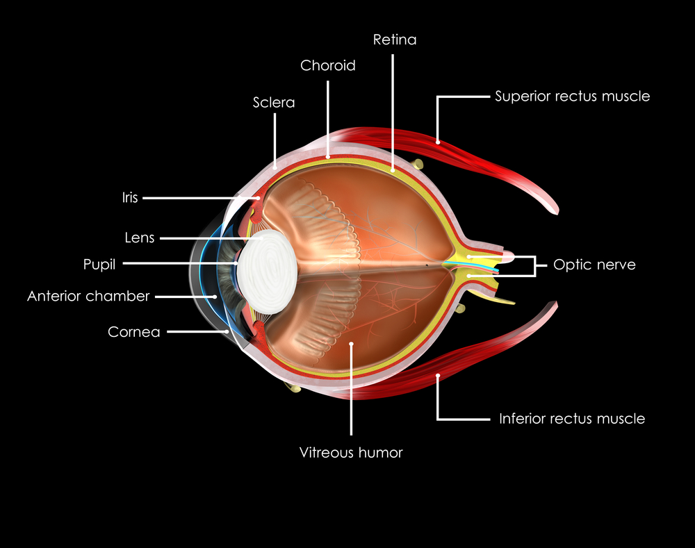
The eye may be small, but it is one of the most amazing parts of your body. To better understand it, it helps to understand the different parts and what they do.

Choroid
A layer with blood vessels that lines the back of the eye and is between the retina (the inner light-sensitive layer that acts like film) and the sclera (the outer white part of the eyeball).

Ciliary Body
The muscle structure behind the iris, which focuses the lens.

Cornea
The very front of the eye that is clear to help focus light into the eye. Corrective laser surgery reshapes the cornea, changing the focus to increase sharpness and/or clarity.

Fovea
The center of the macula which provides the sharp vision.

Iris
The colored part of the eye used to regulate the amount of light entering the eye. Lens focuses light rays onto the retina at the back of the eye. The lens is transparent, and can deteriorate as we age, resulting in the need for reading glasses. Intraocular lenses are used to replace lenses clouded by cataracts.

Macula
The area in the center of retina that contains special light-sensitive cells, allowing us to see fine details clearly in the center of our visual field. The deterioration of the macula can be common as we age, resulting in age related macular degeneration.

Optic Nerve
A bundle of more than a million nerve fibers carrying visual messages from the retina to the brain. Your brain actually controls what you see, since it combines images. Also the images focused on the retina are upside down, so the brain turns images right side up. This reversal of the images Is a lot like what a mirror does in a camera. Glaucoma can result when increase pressure in the eye restricts the flow of impulses to the brain, causing optic nerve damage and makes it difficult to see.

Pupil
The dark center opening in the middle of the iris changes size to adjust for the amount of light available to focus on the retina.

Retina
The nerve layer lining the back of the eye that senses light and creates electrical impulses that are sent through the optic nerve to the brain.

Sclera
The white outer coating of the eyeball.

Vitreous Humor
The clear, gelatinous substance filling the central cavity of the eye.

3/3/16

Susan DeRemerSusan DeRemer, CFRE
Vice President of Development
Discovery Eye Foundation

Pupils Respond To More Than Light

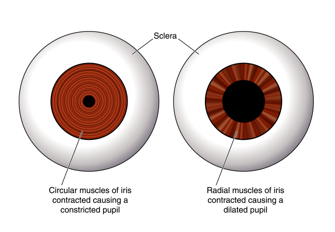
Everyone knows that your pupils will change size according to the amount of light you are experiencing. With more light, the pupil constricts and becomes smaller. Less light and your pupil dilates, letting more light into the back of the eye. It is the muscles of the iris working with your autonomic nervous system (ANS) to adjust the iris so the right amount of light enters the eye – like the aperture of a camera.

The iris is made up of two types of muscle:

  • Sphincter muscles that are like concentric rings that constrict the pupil to as small as two millimeters across
  • Dilator muscles that are laid out like the spokes of a bicycle wheel and can expand the pupil up to eight millimeters across

dilated pupils respond
But the ANS is not only concerned with light reflex, it also reveals emotional and mental responses. The sympathetic branch of the ANS responds to a person being under stress, triggering the “fight or flight” response, which will cause the pupil to dilate. On the other hand, the parasympathetic branch known for “rest and digest” will cause pupil constriction. At any given time, your pupil is balancing between both the light and emotional reactions.

Here are some of examples of mental responses:

Princeton University psychologist Daniel Kahneman demonstrated that pupil size increases in proportion to the difficulty of the task being performed. Calculating 8 x 21 will cause your pupil to dilate slightly, however calculating 8 x 47 will cause them to dilate even more. Whatever the problem, they will remain dilated until you come up with the answer or give up.

Even memory recall creates a pupil response. When subjects were instructed to remember and recite a series of seven digits, their pupils would grow steadily as they learned each number, but reduce as steadily when they recited back each of the numbers.

Wolfgang Einhauser-Treyer, a neurophysicist at Philipps University Marburg in Germany, found that “pupil dilation can betray an individual’s decision before it is openly revealed.” He asked people to push a button at any point during a span of 10 seconds. Dilation began about one second before they pressed the button and continued to peak one to two seconds after the push.

This study of pupil size is known as pupillometry and is used to investigate a wide range of psychological phenomena including sleepiness, introversion, sexual interest, racial bias, schizophrenia, moral judgment, autism and depression. Kahneman said he has “never done any work in which the measurement is so precise.” And while “nobody really knows for sure what these changes do,” according Stuart Steinhauer, director of Biometric Research Lab at the University of Pittsburgh, pupillometry is a valuable tool for psychological research.

So the next time you look into someone’s eyes, know that you have the potential to see more than just their eye color. You might have a clue as to what is going on in their mind.

2/3/15


Susan DeRemerSusan DeRemer, CFRE
Vice President of Development
Discovery Eye Foundation

The Way Eyes Work

9/16/14

Eyes are an amazing part of your body and not just because of what they do helping you see. The are also fascinating be because of the way eyes work. Here are 20 facts about how your eyes function.
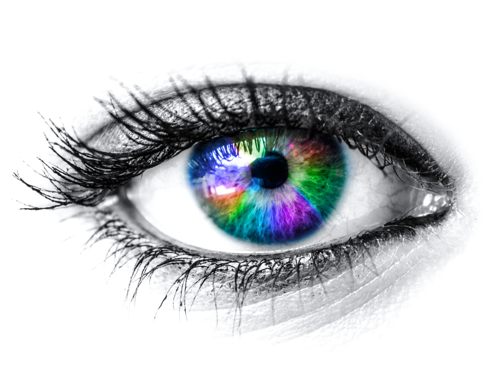
Colorful eye - the way eyes work

      1. The pupil dilates 45% when looking at something pleasant.

2. An eye’s lens is quicker than a camera’s.

3. Each eye contains 107 million cells that are light sensitive.

4. The light sensitivity of rod cells is about 1,000 times that of cone cells.

5. While it takes some time for most parts of your body to warm up their full potential, your eyes are always active.

6. Each of your eyes has a small blind spot in the back of the retina where the optic nerve attaches. You don’t notice the hole in your vision because your eyes work together to fill in each other’s blind spot.

7. The human eye can only make smooth motions if it’s actually tracking a moving object.

8. People generally read 25% slower from a computer screen compared to paper.

9. The eyes can process about 36,000 bits of information each hour.

10. Your eye will focus on about 50 things per second.

11. Eyes use about 65% or your brainpower – more than any other part of your body.

12. Images that are sent to your brain are actually backwards and upside down.

13. Your brain has to interpret the signals your eyes send in order for you to see. Optical illusions occur when your eyes and brain can’t agree.optical illusion - the way eyes work

14. Your pupils can change in diameter from 1 to 8 millimeters, about the size of a chickpea.

15. You see with your brain, not your eyes. Our eyes function like a camera, capturing light and sending data back to the brain.

16. We have two eyeballs in order to give us depth perception – comparing two images allows us to determine how far away an object is from us.

17. It is reported that men can read fine print better than women can.

18. The muscles in the eye are 100 times stronger than they need to be to perform their function.

19. Everyone has one eye that is slightly stronger than the other.

20. In the right conditions and lighting, humans can see the light of a candle from 14 miles away.

Susan DeRemerSusan DeRemer. CFRE
Vice President of Development
Discovery Eye Foundation