Cataract Surgery and Keratoconus

1/8/15

The eye works like a camera, specifically a digital camera. There is the front lens of the camera (cornea), the aperture (iris), the film (retina), and a cable to take the image to the brain (optic nerve). This “camera” also has an additional lens – the natural crystalline lens, which lies behind iris. This natural lens is flexible when we are young, allowing us to focus at distance then instantaneously up close. Around age 40-45, this natural lens starts to stiffen, necessitating the need for reading glasses for most people. This stiffening is the beginning of the aging process that eventually leads to formation of a cataract. We refer to the lens as a cataract when it becomes sufficiently cloudy to affect ones quality of vision.cataract surgery and keratoconus-Cataract diagram 
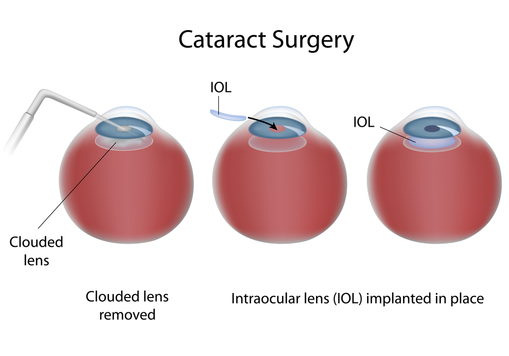
In general, cataract surgery is one of the safest and most successful of all surgeries performed. The basics of cataract surgery in eyes with keratoconus is very similar to non-keratoconic eyes.

Keratoconus (KC) affects this “camera” by causing the front lens (cornea) to bulge. This causes the optics to be distorted. In many cases, this can be corrected for with hard contact lenses (CL) or spectacles; in other cases a corneal transplant may be necessary. When it comes time for cataract surgery in the setting of KC, there are several factors that need to be considered.

Corneal Stability
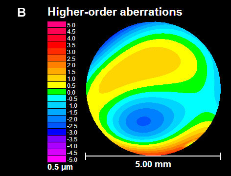
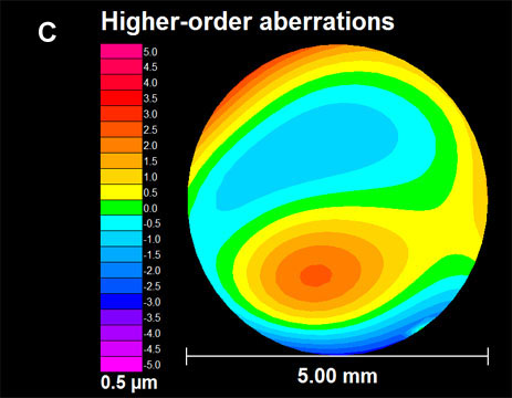
The first thing to be considered is the stability of your cornea. In general, KC progresses more in your late teens to early twenties, and then stabilizes with age. A very exciting treatment for KC is collagen crosslinking. This treatment is meant to stiffen the cornea to prevent instability that is inherent to KC. This treatment promises to stop the progression of KC at a young age. Fortunately, with age, the cornea naturally crosslinks and stiffens, therefore when it comes time for cataract surgery, there is little chance of the progression of KC. Your doctor needs to choose the appropriate intraocular lens (IOL) to refocus your eye after surgery. Two of the most important factors in IOL selection are the length of your eye and the shape of your cornea. Long term CL wear can mold your cornea. It is important to assure that you stay out of your CLs long enough for your cornea to reach its natural shape. Depending on how long you have worn your CLs, it may take several months for the cornea to stabilize. This time can be challenging as your vision will be suboptimal (because you can’t wear CLs), and will be changing (as your cornea reaches its natural shape). When your cornea does stabilize, it is important to determine whether the topography (shape) is regular or irregular. This “regularity” is also known as astigmatism. If the astigmatism is regular, light is focused as a line – generally, this distortion can be fixed with glasses. However, if the astigmatism is irregular, light cannot be focused with glasses, and hard CLs are needed to provide optimal focusing. If you have had a corneal transplant, I generally recommend all your sutures to be removed to allow your new cornea to reach its natural shape.

IOL Selection
The second thing to be considered is the type of IOL. IOLs allow your doctor to refocus the optics of your eye after surgery. In many cases, the correct choice of IOL may decrease your dependence on glasses or CLs. There are several factors that are important when considering the correct IOL for a keratoconic patient. The amount and regularity of your astigmatism plays a very significant role in IOL selection. In general, there are four types of IOLs available in the US – monofocal, toric, pseudo-accomodating, and multifocal. In general I do not recommend multifocal IOLs in patients with KC. These IOLs allow for spectacle independence by spitting the light energy for distance and near, however, with an aberrated cornea (which is what happens in KC), these IOLs do not fare well. If there is a low amount of regular astigmatism or irregular astigmatism, your best bet is a monofocal IOL. This is the “standard” IOL that is covered by your health insurance. If you have higher amounts of astigmatism that your doctor determines is mostly regular, you may benefit from a toric (astigmatism-correcting) IOL. These IOLs can significant improve your uncorrected vision and really decrease your dependence on glasses. It is important to realize that monofocal and toric IOLs only correct vision at one distance. With a monofocal IOL you still can wear a CL to fine-tune your vision, however, with a toric IOL, in general you will need glasses for any residual error. There is a pseudo-accomodating toric IOL available, and this may be a good option if you are trying to decrease your dependence on glasses and correct some of your astigmatism. These IOLs are relatively new to the US market.

If You Had A Corneal Transplant
In the setting of a corneal transplant many of the same factors need to be considered – stability of the graft, choice of IOL, etc. In addition, the health of the graft has to be judged. Prior to cataract surgery in my patients with corneal transplants, I make sure to remove all of their sutures and give the cornea time to stabilize (just as if they were a CTL wearer). If you are a CL wearer, the same rule of being out of the TL until the topography is stable applies. The health of a transplant needs to be established prior to undergoing cataract surgery. The cornea has five main layers to it –cataract surgery and keratoconus-corneal structure 
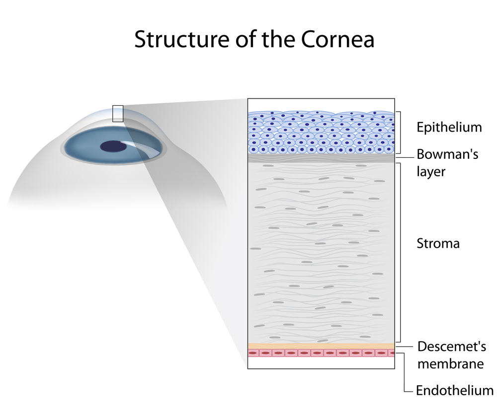
the back layer (inside) is called the endothelium. This layer is responsible for “pumping” fluid out of the cornea, allowing it to stay clear. In all eyes there is a loss of endothelium cells with cataract surgery. I generally perform a “specular microscopy,” which allows me to visualize and quantify the corneal endothelium prior to surgery. This allows me to risk stratify you before your surgery. It is important to realize that corneal transplants have a lifespan and may have to be repeated in the future.

Keep in mind, there is some uncertainty in biometry (the process of selecting an IOL) in all eyes – this error can be higher in keratoconic eyes. This highlights why assuring stability is important. Equally important is picking the correct IOL for your situation. Also, keep in mind that I have discussed generalities in this article. Your individual case could be different. This is a conversation best left between you and your surgeon. In general, cataract surgery and keratoconus or a corneal transplant can be a very safe and effective way in restoring vision.

Sam Garg, MDSumit (Sam) Garg, MD
Interim Chair of Clinical Ophthalmology and Medical Director
Gavin Herbert Eye Institute at the University of California, Irvine

Living With KC Isn’t Easy

10/23/14

The Discovery Eye Foundation Fall 2014 e-newsletter focused on depression and eye disease. At the time we asked for people that were willing to share their stories. Jennifer Villeneuve is one of the many that responded. She is 26 years old and lives in Ontario, Canada. A participant in KC-Link, she talks about the emotional toll keratoconus has taken on her life and living with KC.
Villeneuve with child - living with KC
At age 13, I was diagnosed with keratoconus and given RGP lenses, with which I struggled intensely. ?I became very quiet, and my bubbly personality disappeared. The lenses often got irritated and made my eyes water and turn red, which made me look like I was crying. My doctor didn’t really give me much information on the disease. He just told me my corneas were the shape of footballs instead of circles.

I couldn’t be a normal teenager. I often squinted and had red eyes, which made me very self-conscious. I couldn’t wear makeup or have a free-for-all teenage life. I had to worry about my lenses and what people saw when they looked at me. Some people knew about my KC, but not many. I was just that quiet person who squinted. Because of this, my self-esteem got very low. All in all, my high-school life was hell.

Every time I went to the doctor, he said my pain is normal, the discomfort is typical, and I needed to get used to it. I was also diagnosed with two learning disabilities, in addition to my vision impairment. Throughout high school, I had major anxiety and depression, though I never wanted to admit it. A close teacher even spoke to my mom about the anxiety and low self-esteem. I got through high school, still with the depression being untreated — and still with the same doctor who never even sent me for a topography scan. Each time I went in, it was, “Yup your eyes the same; see ya.”

In college, I was diagnosed with depression and anxiety and was finally getting treated. ?I also went to the University of Ottawa Eye Institute of The Ottawa Hospital in Ontario, Canada. They did topographic scans; my KC had gotten significantly worse. My contacts’ sizing changed twice that year. Again, I couldn’t be normal. College students go out partying, but not me. I stayed in my room by myself. I worried whether I could see enough to go out. What if I drank too much and lost a lens? It wasn’t worth the risk, especially given how expensive they are. In college, I got great help and acceptance. My teachers all knew about my KC, and I was set up with the Centre for Students with Disabilities at Algonquin College in Ottawa. They were my backbone and my support.
Villeneuve with 2 children - living with KC
After my two years in college, I moved on to my career working with children. At each job, I had to explain why I always had a mirror and my contact stuff. At first, I was ashamed and almost embarrassed, explaining why I squinted and that I may not be able to read a kids’ book if the writing is too small. Not only did I get accepted by coworkers, but also by the kids. They knew my eyes were red from my contacts or that my tears meant something was in my eye. I began working in with special-needs children, which was incredible but also challenging — especially in ensuring my eyes were at their best.

At this same time, my vision had gotten worse. I went for corneal crosslinking (CXL) in one eye. I had to take? time off from work, which caused a lot of stress. Unfortunately, there were complications from the CXL. I had a scar in the same eye that caused the crosslinking to be difficult and not as successful. When it came time to do my other eye, I was hesitant, but I needed it. It worked, and the disease slowed down.

A year or so later, the disease had a spike and caused my eye to rub on a lens and make a blister. I had laser surgery to get rid of that and the scar, too. Because KC isn’t covered by insurance in Ontario, I had to pay for every lens, every $11 bottle of solution and countless eye drops. It was expensive, and money was a struggle. I still struggle with the costs of things.

I also have to worry about eye infections. Since I work with kids, they are easy to get, but if I get an eye infection, I can’t wear my contacts, which means no driving, which means no working, which means no money. Things can snowball so quickly.

My vision is up and down. My lenses don’t last as long in my eyes as they used to, and my nighttime vision can be scary. I have to be extra-vigilant. Again, I can’t be normal or go somewhere unfamiliar at night. I’m always concerned. My lens fitter recently recommended scleral lenses, but I can’t afford them. I barely could afford the $2,000 for the CXL.

Living with KC isn’t easy. I can’t help but wonder how long I’ll have the vision I have now. Am I going to be blind in a few years? If I have children, will they have this, or will I even be able to see them? Will I find a guy who would want someone with the possibility of losing vision? I have to stop myself from thinking ahead, or the anxiety gets the best of me.

Janet Villeneuve - living with KCJennifer Villeneuve
Keratoconus Advocate

Children Can Wear Contact Lenses Too

7/24/14

Several studies have shown that children as young as eight years are able to wear gas permeable,[1-3] corneal reshaping,[4-8] and soft contact lenses.[9-12] Gas permeable (hard) contact lenses were originally thought to slow the progression of nearsightedness, but two randomized clinical trials have shown that they do not slow the growth of the eye,[1, 3] so they are primarily fit on children who have difficulty handling soft contact lenses or who have highly irregular corneas (the clear window on the front of the eye) possibly from trauma or eye problems such as keratoconus. children - contact lens for childrenCorneal reshaping contact lenses are worn during sleep only. They temporarily flatten the cornea so that a nearsighted child can see clearly throughout the day with glasses or contact lenses. These contact lenses have been shown to slow eye growth in children.[4-7] Typical soft contact lenses have no effect on the progression of nearsightedness,[13] but soft bifocal contact lenses (typically worn by adults over the age of 40 who otherwise have difficulty seeing clearly at near) have been shown to slow the growth of the eye.[14-17]

Children also benefit from contact lens wear other than slowing the progression of nearsightedness. Children feel better about their athletic abilities, their appearance, and their peer interactions when they wear contact lenses than when they wear spectacles.[11] They even feel smarter if they wear contact lenses than if they wear spectacles, but only if they originally didn’t like to wear spectacles. Children also report that they prefer to participate in activities while wearing contact lenses more than while wearing spectacles, and the most-preferred vision correction is contact lens wear.

It has even been shown that most children (8-12 years of age) require only about five extra minutes to learn how to insert, remove, and care for their contact lenses when compared to teenagers (13-17 years of age). They also show similar benefits as the older group. In fact, children between the ages of 8 and 18 years of age are less likely to require discontinuation of contact lens wear due to problems encountered and also less likely due to experience irritation of the eye due to contact lens wear than college students between the ages of 19 and 25 years.[18, 19] After wearing soft contact lenses for 10 years, those fit as children (7 to 12 years of age) reported similar rates of painful red eyes that required visits to the eye doctor than those fit as teenagers (13 to 17 years of age), and those fit as children as exhibited similar eye health as those fit as teenagers.[20]

Personal experience, backed up by scientific evidence, shows that children as young as eight years can routinely wear contact lenses. When considering contact lens wear for your child, determine the primary reason you would like your child to wear contact lenses. If it is to slow the progression of nearsightedness, then corneal reshaping and soft bifocal contact lenses are the most effective methods. Unfortunately, neither of these contact lenses comes in a daily disposable modality. If your child doesn’t like to wear glasses or finds it difficult to participate in recreational activities with glasses, then daily disposable contact lenses may be best for your child. Contact lenses that are thrown away daily eliminate the need to clean and care for the lenses, reducing care of the lenses to insertion in the morning and removal at bedtime.

Some doctors believe that children should not be fit with contact lenses until they are teenagers. However, there is considerable evidence that indicates children are very capable of contact lens wear, and they experience significant benefits, visually and socially. Talk to your eye doctor about contact lens wear for your child, and if your doctor says that children should not be fit with contact lenses, consider a second opinion.

References
[1] Katz J, Schein OD, Levy B, et al. A randomized trial of rigid gas permeable contact lenses to reduce progression of children’s myopia. Am J Ophthalmol 2003;136:82-90. (Go Back)
[2] Khoo CY, Chong J, Rajan U. A 3-year study on the effect of RGP contact lenses on myopic children. Singapore Med J 1999;40:230-7. (Go Back)
[3] Walline JJ, Jones LA, Mutti DO, et al. A randomized trial of the effects of rigid contact lenses on myopia progression. Arch Ophthalmol 2004;122:1760-6. (Go Back)
[4] Cho P, Cheung SW. Retardation of Myopia in Orthokeratology (ROMIO) Study: A 2-Year Randomized Clinical Trial. Invest Ophthalmol Vis Sci 2012;53:7077-85. (Go Back)
[5] Cho P, Cheung SW, Edwards M. The longitudinal orthokeratology research in children (LORIC) in Hong Kong: a pilot study on refractive changes and myopic control. Curr Eye Res 2005;30:71-80. (Go Back)
[6] Santodomingo-Rubido J, Villa-Collar C, Gilmartin B, et al. Myopia Control with Orthokeratology Contact Lenses in Spain (MCOS): Refractive and Biometric Changes. Invest Ophthalmol Vis Sci 2012. (Go Back)
[7] Walline JJ, Jones LA, Sinnott LT. Corneal reshaping and myopia progression. Br J Ophthalmol 2009;93:1181-5. (Go Back)
[8] Walline JJ, Rah MJ, Jones LA. The Children’s Overnight Orthokeratology Investigation (COOKI) pilot study. Optom Vis Sci 2004;81:407-13. (Go Back)
[9] Rah MJ, Walline JJ, Jones-Jordan LA, et al. Vision specific quality of life of pediatric contact lens wearers. Optom Vis Sci 2010;87:560-6. (Go Back)
[10] Walline JJ, Gaume A, Jones LA, et al. Benefits of Contact Lens Wear for Children and Teens. Eye Contact Lens 2007;33:317-21. (Go Back)
[11] Walline JJ, Jones LA, Sinnott L, et al. Randomized trial of the effect of contact lens wear on self-perception in children. Optom Vis Sci 2009;86:222-32. (Go Back)
[12] Walline JJ, Long S, Zadnik K. Daily disposable contact lens wear in myopic children. Optom Vis Sci 2004;81:255-9. (Go Back)
[13] Walline JJ, Jones LA, Sinnott L, et al. A randomized trial of the effect of soft contact lenses on myopia progression in children. Invest Ophthalmol Vis Sci 2008;49:4702-6. (Go Back)
[14] Anstice NS, Phillips JR. Effect of dual-focus soft contact lens wear on axial myopia progression in children. Ophthalmology 2011;118:1152-61. (Go Back)
[15] Lam CS, Tang WC, Tse DY, et al. Defocus Incorporated Soft Contact (DISC) lens slows myopia progression in Hong Kong Chinese schoolchildren: a 2-year randomised clinical trial. Br J Ophthalmol 2014;98:40-5. (Go Back)
[16] Sankaridurg P, Holden B, Smith E, 3rd, et al. Decrease in rate of myopia progression with a contact lens designed to reduce relative peripheral hyperopia: one-year results. Invest Ophthalmol Vis Sci 2011;52:9362-7. (Go Back)
[17] Walline JJ, Greiner KL, McVey ME, et al. Multifocal contact lens myopia control. Optom Vis Sci 2013;90:1207-14. (Go Back)
[18] Wagner H, Chalmers RL, Mitchell GL, et al. Risk Factors for Interruption to Soft Contact Lens Wear in Children and Young Adults. Optom Vis Sci 2011;88:973-80. (Go Back)
[19] Wagner H, Richdale K, Mitchell GL, et al. Age, behavior, environment, and health factors in the soft contact lens risk survey. Optom Vis Sci 2014;91:252-61. (Go Back)
[20] Walline JJ, Lorenz KO, Nichols JJ. Long-term contact lens wear of children and teens. Eye Contact Lens 2013;39:283-9. (Go Back)

Jeffrey Walline - contact lenses childernJeffrey J. Walline, OD, PhD
Associate Professor
Chair, Research and Graduate Studies
The Ohio State University College of Optometry

Breathing, Patience and Keratoconus

7/1/14

Besides hearing from eye care professionals and Discovery Eye Foundation staff, we also think it is important to hear from people with sight threatening eye diseases such as age-related macular degeneration, keratoconus, retinitis pigmentosa, etc. They can share their experiences with others that are newly diagnosed with eye disease, while providing insights to family members, friends and caregivers. What follows is the first of these occasional posts focusing on the experiences and insights gained from vision loss.

“The expression on my optometrist’s face was as if he was giving me a cancer sentence,” Jeanette Hasseman remembers. “I had never heard of keratoconus, but he told me it was an incurable vision disability. He said he was aware of some evolving technology, but he wanted to give me some time to absorb and research the disease.
Jeanette Hasseman - keratoconus
“When I went back three months later, he told me a about a cross-linking study that was going on not-too-far from here, but I hadn’t fully absorbed all the information I was reading yet,” she recalls. I did not understand — even though it was plainly stated in the information I was looking at — that once you lose some vision, you don’t get it back.

“I kept thinking, ‘OK, this certainly can be fixed,” so instead of getting cross-linking in July, I waited until December. I regret not acting on my doctor’s suggestion of getting in the study in July, because I lost a tremendous amount of vision by December.”

Hasseman was tested and qualified for the clinical trial in December and had epi-on cross-linking in both eyes in January 2013. “The topography scans show the KC progression has been halted,” she says. “I rejoice in that.”

While her vision seemed better shortly after the procedures, she was diagnosed one week after crosslinking with a “gouge” (4 mm corneal abrasion) in her left cornea that resulted in severe hazing. While the severity of the corneal hazing has decreased in the past year, she is waiting to find out if it is permanent. If so, she may need to have a corneal transplant at some point to restore any visual acuity.

A registered nurse, Hasseman finished her BSN degree two years ago at age 54 — just months before she was diagnosed with KC. The native Ohioan has since stopped working as a nurse, and more recently, she had to give up teaching and doing tatting — lace-making — a hobby she had enjoyed for decades.

Hasseman found the National Keratoconus Foundation (NKCF) when she first Googled “kerotoconus.” “KC-Link has been a great blessing,” she says. “I’ve downloaded information; I signed up for KC-Link and asked questions of Catherine [Warren, director of NKCF] and the moderating doctors. I found great support for my own spirit, as well as information on the latest technologies.

“When people ask questions on KC-Link, if I can relate, I answer. Just the other day, someone who was just diagnosed asked, ‘Well, what should I do?’ I wrote: ‘First thing is: Breathe. Second thing is: Ask for information from your eye doctor and ask who is the best corneal specialist in your area who is really good for keratoconus. Most of all: Be patient.’

“It’s really hard to lose your vision. Even in your own heart, you can feel isolated. KC-Link gives you a body of other people who totally understand what you’re thinking, what you’re feeling, what frustrates you. There is so much advice you can get on KC-Link regarding contacts, how to keep eyes moist, how to handle low-light driving, what type of e-reader works best for people with keratoconus — you cannot exhaust the information that is shared; you cannot exhaust the different emotions that are shared.”

Jeanette HassemanJeanette Hasseman
Keratoconus Advocate

New Technology for Evaluating Contact Lenses

5/1/14

Successful management with contact lenses can sometimes be a frustrating process for those with keratoconus. The fitting and evaluation process involves numerous visits to the optometrist, out of pocket expenses as well as medical insurance co-pays. Luckily, the contact lens industry has responded to the need to have better contact lens materials for patients with keratoconus. It used to be that the only contact lenses available to manage keratoconus were gas-permeable (GP) lenses. Now, more and more patients are being fit with newer generation hybrid lenses (GP lens core with a skirt of soft lens material to aid in fit and comfort) and scleral lenses (large diameter GP lenses that do not rest on the cornea, only the outlying sclera). These newer designs are intended to vault over the central cornea and do not rest on the cornea at all. They have resulted in much more comfortable and wearable strategies for full-time use. As a result of the newer lens designs, the game has changed when it comes to the science of fitting and evaluating the lenses.

For years, optometrists have used corneal topography to guide their decision making on fitting keratoconic eyes. A corneal topographer is an instrument that maps the shape of the cornea, and gives information much like that of a topographical map for hiking. Corneal topography is still an absolutely mandatory part of evaluating the shape of the cone, the simulated corneal curvature, and monitoring for progression of the disease, and is not a standard part of a typical eye examination. However, doctors have a new tool at their disposal for fitting contact lenses on patients with keratoconus.

Optical Coherence Tomography (OCT) was once reserved for use in the back of the eye, or retina. OCT uses visible light passed through the clear structures of the eye to generate a cross-sectional image of the layers of the retina, much like an image generated by an MRI. Advances in OCT technology has improved the resolution to image the eye on the micrometer scale (one-thousandth of a millimeter). OCT technology is now commercially available not only for the retina, but the structures of the front part of the eye. The obvious application is to aid the doctor in the fitting and evaluating complex contact lenses that vault the cornea.

Figure 1.  OCT image of a scleral lens fit on a keratoconic patient.  The cornea is the opaque white band located at the bottom of this picture, the tear film reservoir is the middle clear band and the contact lens is the top band.  Using an electronic caliper tool, the precise amount of vault can be measured, leaving no doubt as to the precision of the fit.
Figure 1. OCT image of a scleral lens fit on a keratoconic patient. The cornea is the opaque white band located at the bottom of this picture, the tear film reservoir is the middle clear band and the contact lens is the top band. Using an electronic caliper tool, the precise amount of vault can be measured, leaving no doubt as to the precision of the fit.

OCT allows the optometrist to view a cross-sectional image of the contact lens on the eye in real time and to monitor the health of the cornea in the presence of the contact lens. This view is valuable for judging the vault of new designs of contact lenses over the cornea and judging where the lenses land on the eye. It is the most specific way to determine if the fit is acceptable and to troubleshoot if lenses are not fitting appropriately.

Figure 2.  OCT image of the periphery of a scleral lens on a patient with pellucid marginal degeneration.  The lens contacts the cornea over an area of 0.87mm long.  These types of measurements help guide decision making in modifying the lens fit and were impossible before the advent of this technology.
Figure 2. OCT image of the periphery of a scleral lens on a patient with pellucid marginal degeneration. The lens contacts the cornea over an area of 0.87mm long. These types of measurements help guide decision making in modifying the lens fit and were impossible before the advent of this technology.
Figure 3.  Hybrid lens on a highly irregular eye after corneal transplant.  The point of contact of the soft skirt with the cornea is visible to the right of the image.
Figure 3. Hybrid lens on a highly irregular eye after corneal transplant. The point of contact of the soft skirt with the cornea is visible to the right of the image.

Optometrists now have a much more powerful tool for evaluating and managing even the most challenging contact lens fits. It remains to be seen whether this technology has the ability to reduce the number of visits required for successful fit. But, the precision afforded by this technology does have the ability to improve patient outcomes.

Sonsino HeadshotJeffrey Sonsino, OD, FAAO
The Contact Lens Center at Optique Diplomate
Cornea, Contact Lens, and Refractive Therapies

Wavefront Sensing Applied to Custom Contact Lens Research in Keratoconus

4/10/14

During a trip to the optometrist or ophthalmologist, a patient will encounter the process of subjective refraction.  This technique involves the clinician asking the patient to make a series of judgments (which is better, one or two?) about the clarity of their vision when looking through a series of lenses.  The choices that the patient makes guide the clinician in identifying an optical prescription which is typically made up of sphere, and potentially, cylinder lenses.

Why is it that glasses don’t always work for patients with keratoconus?

In many instances, individuals with keratoconus do not achieve excellent visual performance with spectacles or traditional soft contact lenses.  One cause for the failure of these corrections is that the changes in corneal shape that accompany keratoconus induce refractive errors which traditional spectacles simply cannot correct.  So, even when sphere and cylinder in the keratoconic eye are well-corrected, these “other refractive errors” or “other aberrations” remain uncorrected and can lead to a blurred retinal image and blurred vision.  Collectively these other aberrations can be referred to as higher order aberration, while the aberrations that are typically corrected with spectacles and soft contact lenses are referred to as lower order aberration.

What kinds of higher order aberrations are present in keratoconus:

Pantanelli et al. have stated that the level of higher order aberration present in an eye with keratoconus is, on average, approximately 5.5 times higher than the level experienced in a control group.  In an effort to visualize higher-order aberration data, they are commonly represented graphically as shown in the figures below.  Examples of higher order aberration measured in one normal eye are shown in figure A, while an example of higher order aberration from one keratoconic eye are shown in figure B.  The circular nature of the map denotes the boundary of the measurement, which is defined by the round pupil of the eye.  A majority of the higher order aberration map in figure A is green (denoting a relative absence of higher order aberration).  However, the map in figure B displays a much larger variation in color, indicating the presence of higher order aberration  in this individual keratoconic eye in a greater quantity than the normal eye shown in figure A.

Figure A - normal-keratoconus
Figure A – normal
Figure B - keratoconus
Figure B – keratoconus

A wavefront aberration map of the “other aberrations” or higher order aberrations of two eyes. Figure A is an example of data for a normal eye and figure B is an example of data for an eye with keratoconus.

If refraction is not capable of quantifying higher order aberrations, how are they measured?

One method for obtaining the information regarding higher order aberration shown above is with a wavefront sensor.  The wavefront sensor objectively (without patient feedback) collects information on the optical performance of the eye that can be used to calculate the amount of both lower and higher order aberration present.

Laboratory-based research related to custom contact lenses:

Several investigators in the laboratory (e.g. Katsoulos et al., Sabesan et al., Chen et al., Marsack et al.) have reported on work that attempts to further reduce higher order aberration by targeting the eye-specific higher order aberration seen in a given keratoconic eye.  The general philosophy behind these customized lenses is that the aberration pattern measured with the wavefront sensor is a more complete optical prescription for implementation of a custom contact lens.  Figure C demonstrates, in principle, the optical properties of a contact lens designed to correct the higher-order aberration in figure B.  Where the map of the eye (figure B) is red, the map of the correction (figure C) is blue, and vice versa.  When the lens is worn, the net effect as light propagates through the lens-eye system is the cancellation of the higher order aberration in a targeted manner.

Figure C -keratoconus correction

In principle, this figure pictorially represents the higher order optical properties of a contact lens designed to fully correct the higher-order aberration of the eye represented in figure B.

What is next:

Investigators continue to push the technology behind custom contact lenses for keratoconus towards clinical relevance.  However, like every novel intervention strategy, we must manage our expectations.  Complexity in measuring keratoconic eyes, a need for specialized equipment and expertise to design and manufacture the lenses, the infrastructure needed to coordinate the clinical exam and manufacture efforts and cost associated with the process are a subset of the barriers that must be removed if this type of correction is to become more mainstream.  For this reason, it is my opinion that if/when these corrections become commonly available in the clinic, they will likely add to, and not replace, existing forms of corrections that patients and clinicians now utilize to correct vision.

jmarsack-bio-picJason Marsack, PhD
Research Assistant Professor
University of Houston, College of Optometry.
Dr. Marsack’s work focuses on the relationship between visual performance
and optical aberration in individuals with highly aberrated eyes.

Help for Computer Users

Working long hours in front of the computer requires a fairly unchanging body, head and eye position which can cause discomfort.  Correct working position, periodic stretch breaks, frequent eye blinking, artificial tears for lubrication are all very important.  However, it’s not always easy to remember this when you are engrossed in work. Here are a few fun, free and easy-to-install “break reminders” to help:

WorkSafe Sam - break reminder
WorkSafe Sam
WorkSafe Sam is a desktop tool that provides stretching tips to help reduce eye and muscle strain for office workers (clicking on this link will open a file on your computer because this is a zip file).

Workrave is break reminder program that alerts you to take “micro-pauses” and stretch breaks.

Take Your Break is another break reminder designed to prevent or minimize repetitive strain injury, computer eye strain and other computer related health problems.  It has a friendly interface and a tray icon status indicator.  It runs quietly in the background, monitoring your activity and reminding you to take regular breaks.

And remember to blink.  Blinking cleans the ocular surface of debris and flushes fresh tears over the ocular surface. Each blink brings nutrients to the eye surface structures keeping them healthy. The flow of tears is responsible for wetting the lower third of the cornea. This is very important in KC, since this area is generally below the bulge of the cone and in many cases irritated by wobbly RGP lenses.  Maybe your job requires hours of work at a computer. Maybe you like to spend your free time surfing the internet. Whatever the reason, your body is probably feeling the effects of spending too much time staring at a computer monitor, which could result in Computer Vision Syndrome (CVS).  The most common symptoms are: eye strain, dry or irritated eyes,redness in eyes,difficulty in refocusing eye,neck pain,double vision,blurred vision, fatigue, and headaches.

Please join us on Thursday when Dr. Bezalel Schendowich will be providing a detailed insight into the importance of blinking, going beyond computer usage.

CathyW headshotCathy Warren, RN
Executive Director
National Keratoconus Foundation

Corneal Transplant Surgery Options

In this day and age of advancing technology, corneal transplants have changed from a long arduous ordeal to a more simple and precise procedure that offers faster visual recovery.  Instead of replacing the entire cornea for any and all corneal diseases, we now perform disease targeted partial corneal transplants.  If the disease involves the back layer of the cornea, we perform endothelial keratoplasty and replace only the diseased inner layer of the cornea.  Conversely, if the problematic portions are the front layers of the cornea, we perform anterior lamellar keratoplasty.  The co-morbidity and risk of rejection from partial corneal transplants are significantly less than the traditional full thickness transplants.

With endothelial keratoplasty, a small incision, about 4-5 mm is made and a sheet of donor endothelial cells are placed into the anterior chamber of the eye.  A large air bubble is then used to float this sheet up so that it opposes the posterior or back portion of the cornea.  The patient is asked to position face up for 24 hours.  Over this period of time, the cells will “stick” on their own and thus no sutures are required to keep the graft in place.,/span>

Figure 1 - corneal transplant
Figure 1

Anterior lamellar keratoplasty is done for superficial scars and opacities of the cornea or for keratoconus, a genetic degeneration of the cornea that is seen in younger individuals.  In this case, the native endothelial cells of the patient are healthy and therefore are left intact while the remainder of the cornea is transplanted.  This significantly lowers the risk of rejection, which is traditionally a much higher risk in young patients.  Multiple sutures are required to maintain this graft in place however, with the advent of femtosecond laser technology, the wound configuration is made in such a way as to promote rapid healing and visual recovery. (Figure 1)  Sutures are removed at an earlier time than with traditional surgery and the eye is able to undergo visual rehabilitation with glasses or contact lenses in 3-6 months’ time.

Corneal transplantation does not require waiting on a list for a donor to become available like it once did.  There are now multiple excellent eye banks across America that harvest, screen, and distribute donor tissue to surgeons.  This way, tissue is readily available and patients only need to schedule a time based on their own and their surgeon’s time schedule.  Post operatively, patients are asked to return to regular activity with the exception of no heavy lifting or bending for a period of 2 months.  Antibiotic and anti-rejection drops are started immediately after surgery and continued for several months after.  No oral medications aside from the patient’s regular medications are required.

Farid 3.6.14Marjan Farid, MD
Director of Cornea, Cataract, and Refractive Surgery
Vice-Chair of Ophthalmic Faculty
Director of the Cornea Fellowship Program
Associate Professor of Ophthalmology
Gavin Herbert Eye Institute, University of California, Irvine