When Is The Best Time For Cataract Surgery?

As you age, cataracts become a concern prompting the question – when is the best time for cataract surgery?

There are decades worth of old wives tales floating around regarding cataracts that often lead to unnecessary fear and apprehension for many patients. These myths involve concepts such as “ripeness”, having to wear eye patches afterwards, danger in “waiting too long, etc. Just as the techniques of cataract extraction have changed over the decades, so have the indications to proceed to surgery.
best time for cataract surgery - people
Firstly, cataracts are a normal part of the aging process. Patients should not be alarmed if they are told that they are developing cataracts, even as early as their fifties. As we age, the natural clear lens inside the eye becomes progressively harder, darker, and cloudier. This dark, cloudy lens is what is referred to as a cataract. Cataracts develop at different rates for different people, and even between the two eyes of the same person. It typically takes many years for the lens to become cloudy enough to impact the clarity of vision. There are many different types of cataracts depending of what area of the lens becomes cloudy, but the typical cataract related to normal aging results in a relatively uniform cloudiness with a denser central core, and is referred to as “Nuclear Sclerosis”. Other varieties of cataracts tend to grow more quickly, are relatively uncommon, and often result from certain conditions other than typical aging.
best time for cataract surgery
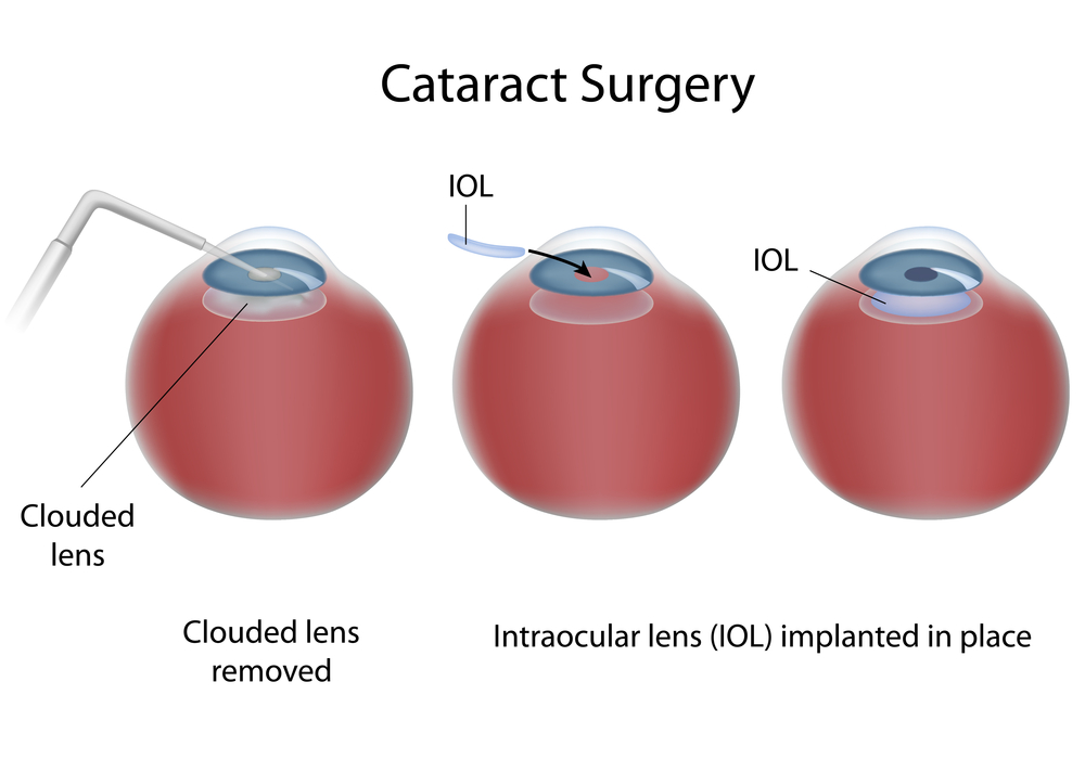
Regardless of what type of cataract the patient has, the treatment is the same: cataract extraction with an implant of an intraocular lens. There have been great advances in lens design over the years, and they now result in excellent, stable, predictable vision for the remainder of the patient’s lifetime and do not typically need to be changed once implanted.

Cataracts result in different symptoms that may be more of less relevant to a specific person’s needs, such as:

  • Glare with bright lights
  • Difficulty with fine print
  • Difficulty following the golf or tennis ball
  • Impairment in night driving
  • Difficulty with seeing street signs
  • Seeing the score or small print on the television
  • Fine visual tasks such as threading a needle, etc.

Although cataract surgery is an incredibly successful procedure with only about a 1-2% risk of complications, it still DOES have some risk. Therefore, cataract surgery should only be undertaken when there is something to gain. In other words, the BENEFITS MUST OUTWEIGH THE RISKS. This means that if your symptoms are mild and are not interfering with your activities of daily living, it is not time to accept the risks of surgery. Once your visual impairment progresses to the point that YOU feel your activities of daily living and enjoyment are impaired, this is the time to proceed to surgery. This threshold is very different between people. Some people feel impaired with vision of 20/25, and others still function within their scope of usual activities until they are 20/100! The best first-step in determining if it is time for your surgery is to get an up-to date refraction. This means a detailed check for new glasses. Often, cataract development will change a person’s glasses prescription, and updating this can improve the visual symptoms for months to years. When a new glasses prescription no longer improves the sight adequately, this is when surgery is indicated.

For the most part, putting off cataract surgery does not impact the final outcome. It will not harm you or your eye to leave the cataract alone until you are ready. There are of course certain exceptions to this rule, such as in Fuchs’ dystrophy, pseudoexfolation, untreated narrow-angle glaucoma, and some others. However, these are relatively rare conditions that your doctor will speak to you about if you have any of these diagnoses.

In summary, the time to proceed to cataract surgery is something that you as the patient determine. YOU assess your lifestyle needs and your vision performance within your scope of activities. When you feel you are impaired in these activities, the benefits will outweigh the risks, and it’s time to take them out. You should not feel any pressure to urgency in this process.

Once you have determined you are ready to have cataract surgery, your surgeon will discuss with you your options for intraocular lens implantation including astigmatism neutralizing lenses, standard distance or near-vision lenses, multiple focal distance lenses, accommodating lenses, and others. The current standard approach for cataract surgery is called “phacoemulsification” and uses ultrasound technology to remove the cataract. There are also laser devices that assist in making the incisions and breaking up the lens, which many surgeons now employ in addition to the phacoemulsification. In general cataract surgery only takes a few minutes, is performed with topical anesthesia, is pain-free, and has a very short recovery time. No pirate-patches are used these days! Most patients are very happy with the results, but this requires adequate discussion with the surgeon prior to the procedure to best assess the needs of the individual patient. A well- informed patient who participates in their care results in the best outcomes!

6/18/15

Sameh Mosaed, MD best time for cataract surgerySameh Mosaed, MD
Director of Glaucoma Services, Gavin Herbert Eye Institute, UC Irvine
Associate Professor, Cataract and Glaucoma Surgery, UC Irvine School of Medicine

Cataract Surgery and Keratoconus

1/8/15

The eye works like a camera, specifically a digital camera. There is the front lens of the camera (cornea), the aperture (iris), the film (retina), and a cable to take the image to the brain (optic nerve). This “camera” also has an additional lens – the natural crystalline lens, which lies behind iris. This natural lens is flexible when we are young, allowing us to focus at distance then instantaneously up close. Around age 40-45, this natural lens starts to stiffen, necessitating the need for reading glasses for most people. This stiffening is the beginning of the aging process that eventually leads to formation of a cataract. We refer to the lens as a cataract when it becomes sufficiently cloudy to affect ones quality of vision.cataract surgery and keratoconus-Cataract diagram In general, cataract surgery is one of the safest and most successful of all surgeries performed. The basics of cataract surgery in eyes with keratoconus is very similar to non-keratoconic eyes.

Keratoconus (KC) affects this “camera” by causing the front lens (cornea) to bulge. This causes the optics to be distorted. In many cases, this can be corrected for with hard contact lenses (CL) or spectacles; in other cases a corneal transplant may be necessary. When it comes time for cataract surgery in the setting of KC, there are several factors that need to be considered.

Corneal Stability
The first thing to be considered is the stability of your cornea. In general, KC progresses more in your late teens to early twenties, and then stabilizes with age. A very exciting treatment for KC is collagen crosslinking. This treatment is meant to stiffen the cornea to prevent instability that is inherent to KC. This treatment promises to stop the progression of KC at a young age. Fortunately, with age, the cornea naturally crosslinks and stiffens, therefore when it comes time for cataract surgery, there is little chance of the progression of KC. Your doctor needs to choose the appropriate intraocular lens (IOL) to refocus your eye after surgery. Two of the most important factors in IOL selection are the length of your eye and the shape of your cornea. Long term CL wear can mold your cornea. It is important to assure that you stay out of your CLs long enough for your cornea to reach its natural shape. Depending on how long you have worn your CLs, it may take several months for the cornea to stabilize. This time can be challenging as your vision will be suboptimal (because you can’t wear CLs), and will be changing (as your cornea reaches its natural shape). When your cornea does stabilize, it is important to determine whether the topography (shape) is regular or irregular. This “regularity” is also known as astigmatism. If the astigmatism is regular, light is focused as a line – generally, this distortion can be fixed with glasses. However, if the astigmatism is irregular, light cannot be focused with glasses, and hard CLs are needed to provide optimal focusing. If you have had a corneal transplant, I generally recommend all your sutures to be removed to allow your new cornea to reach its natural shape.

IOL Selection
The second thing to be considered is the type of IOL. IOLs allow your doctor to refocus the optics of your eye after surgery. In many cases, the correct choice of IOL may decrease your dependence on glasses or CLs. There are several factors that are important when considering the correct IOL for a keratoconic patient. The amount and regularity of your astigmatism plays a very significant role in IOL selection. In general, there are four types of IOLs available in the US – monofocal, toric, pseudo-accomodating, and multifocal. In general I do not recommend multifocal IOLs in patients with KC. These IOLs allow for spectacle independence by spitting the light energy for distance and near, however, with an aberrated cornea (which is what happens in KC), these IOLs do not fare well. If there is a low amount of regular astigmatism or irregular astigmatism, your best bet is a monofocal IOL. This is the “standard” IOL that is covered by your health insurance. If you have higher amounts of astigmatism that your doctor determines is mostly regular, you may benefit from a toric (astigmatism-correcting) IOL. These IOLs can significant improve your uncorrected vision and really decrease your dependence on glasses. It is important to realize that monofocal and toric IOLs only correct vision at one distance. With a monofocal IOL you still can wear a CL to fine-tune your vision, however, with a toric IOL, in general you will need glasses for any residual error. There is a pseudo-accomodating toric IOL available, and this may be a good option if you are trying to decrease your dependence on glasses and correct some of your astigmatism. These IOLs are relatively new to the US market.

If You Had A Corneal Transplant
In the setting of a corneal transplant many of the same factors need to be considered – stability of the graft, choice of IOL, etc. In addition, the health of the graft has to be judged. Prior to cataract surgery in my patients with corneal transplants, I make sure to remove all of their sutures and give the cornea time to stabilize (just as if they were a CTL wearer). If you are a CL wearer, the same rule of being out of the TL until the topography is stable applies. The health of a transplant needs to be established prior to undergoing cataract surgery. The cornea has five main layers to it –cataract surgery and keratoconus-corneal structure the back layer (inside) is called the endothelium. This layer is responsible for “pumping” fluid out of the cornea, allowing it to stay clear. In all eyes there is a loss of endothelium cells with cataract surgery. I generally perform a “specular microscopy,” which allows me to visualize and quantify the corneal endothelium prior to surgery. This allows me to risk stratify you before your surgery. It is important to realize that corneal transplants have a lifespan and may have to be repeated in the future.

Keep in mind, there is some uncertainty in biometry (the process of selecting an IOL) in all eyes – this error can be higher in keratoconic eyes. This highlights why assuring stability is important. Equally important is picking the correct IOL for your situation. Also, keep in mind that I have discussed generalities in this article. Your individual case could be different. This is a conversation best left between you and your surgeon. In general, cataract surgery and keratoconus or a corneal transplant can be a very safe and effective way in restoring vision.

Sam Garg, MDSumit (Sam) Garg, MD
Interim Chair of Clinical Ophthalmology and Medical Director
Gavin Herbert Eye Institute at the University of California, Irvine