Corneal Donor’s Age Not Critical for Transplant Success

6/17/14

In the United States, more than 40,000 corneal transplants are performed each year with a high success rate in comparison to other types of organ transplants. According to the Eye Bank Association of America (EBAA), keratoconus was the leading cause of anterior lamellar keratoplasty (DALK/ALK partial thickness transplant) and was the fourth most common indication for penetrating keratoplasty surgery in 2012 (their last reporting period).
corneal transplant-original size
Advances in technology have led to increasingly successful outcomes for all who need corneal transplants. New long-term research of corneal transplant patients have shown that the age of corneal donors is no longer as important as once thought by eye health providers. According to a study funded by the National Institutes of Health, ten years after a transplant, a cornea from a 71-year-old donor is likely to remain as healthy as a cornea from a donor half that age.

The Cornea Donor Study (see www.ClinicalTrials.gov), funded by NIH’s National Eye Institute (NEI), was designed to compare graft survival rates for corneas from two donor age groups, aged 12-65 and aged 66-75. It was coordinated by the Jaeb Center for Health Research in Tampa, Fla., and involved 80 clinical sites across the United States. The study enrolled 1,090 people eligible for transplants, ages 40-80. Donor corneas were provided by 43 eye banks, and met the quality standards of the Eye Bank Association of America. The study found that 10-year success rates remained steady at 75 percent for corneal transplants from donors 34-71 years old. In the United States, three-fourths of cornea donors are within this age range, and one-third of donors are at the upper end of the range, from 61-70 years old.

Prior to this study, many surgeons would not accept corneas from donors over 65. Since the supply of young donor corneas is limited, these study results are encouraging for those who face a corneal transplant . The high level of success rates using corneas from older donors (over age 60) greatly increases the pool of donated corneas and corneal tissue available for transplant. In 2012, corneal donors under age 31 comprised less than 10 percent of the U.S. donor pool. “Our study supports continued expansion of the corneal donor pool beyond age 65,” said study co-chair Edward J. Holland, M.D., professor of ophthalmology at the University of Cincinnati and director of the Cornea Service at the Cincinnati Eye Institute. “We found that transplant success rates were similar across a broad range of donor ages.”

“Overall, the findings clearly demonstrate that most corneal transplants have remarkable longevity regardless of donor age,” said Mark Mannis, M.D., chair of ophthalmology at the University of California, Davis, and co-chair of the study. “The majority of patients continued to do well after 10 years, even those who received corneas from the oldest donors.”

SOURCE: National Eye Institute Press Releases

For information about Eye Bank Association of America

CathyW headshotCathy Warren, RN
Executive Director
National Keratoconus Foundation

Corneal Transplant Surgery Options

In this day and age of advancing technology, corneal transplants have changed from a long arduous ordeal to a more simple and precise procedure that offers faster visual recovery.  Instead of replacing the entire cornea for any and all corneal diseases, we now perform disease targeted partial corneal transplants.  If the disease involves the back layer of the cornea, we perform endothelial keratoplasty and replace only the diseased inner layer of the cornea.  Conversely, if the problematic portions are the front layers of the cornea, we perform anterior lamellar keratoplasty.  The co-morbidity and risk of rejection from partial corneal transplants are significantly less than the traditional full thickness transplants.

With endothelial keratoplasty, a small incision, about 4-5 mm is made and a sheet of donor endothelial cells are placed into the anterior chamber of the eye.  A large air bubble is then used to float this sheet up so that it opposes the posterior or back portion of the cornea.  The patient is asked to position face up for 24 hours.  Over this period of time, the cells will “stick” on their own and thus no sutures are required to keep the graft in place.,/span>

Figure 1 - corneal transplant
Figure 1

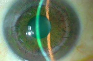
Anterior lamellar keratoplasty is done for superficial scars and opacities of the cornea or for keratoconus, a genetic degeneration of the cornea that is seen in younger individuals.  In this case, the native endothelial cells of the patient are healthy and therefore are left intact while the remainder of the cornea is transplanted.  This significantly lowers the risk of rejection, which is traditionally a much higher risk in young patients.  Multiple sutures are required to maintain this graft in place however, with the advent of femtosecond laser technology, the wound configuration is made in such a way as to promote rapid healing and visual recovery. (Figure 1)  Sutures are removed at an earlier time than with traditional surgery and the eye is able to undergo visual rehabilitation with glasses or contact lenses in 3-6 months’ time.

Corneal transplantation does not require waiting on a list for a donor to become available like it once did.  There are now multiple excellent eye banks across America that harvest, screen, and distribute donor tissue to surgeons.  This way, tissue is readily available and patients only need to schedule a time based on their own and their surgeon’s time schedule.  Post operatively, patients are asked to return to regular activity with the exception of no heavy lifting or bending for a period of 2 months.  Antibiotic and anti-rejection drops are started immediately after surgery and continued for several months after.  No oral medications aside from the patient’s regular medications are required.

Farid 3.6.14Marjan Farid, MD
Director of Cornea, Cataract, and Refractive Surgery
Vice-Chair of Ophthalmic Faculty
Director of the Cornea Fellowship Program
Associate Professor of Ophthalmology
Gavin Herbert Eye Institute, University of California, Irvine