Telemedicine For Diagnosing Retinopathy Of Prematurity

8/28/14

Technology now allows patients access to highly-qualified, specialized care, no matter where they live. This potential is demonstrated in the sight-saving treatments that can be used to help children lead normal healthy lives. The following article is posted with the permission of the National Institute of Health (NIH).

Telemedicine is an effective strategy to screen for the potentially blinding disease known as retinopathy of prematurity, according to a study funded by the National Eye Institute (NEI). The investigators say that the approach, if adopted broadly, could help ease the strain on hospitals with limited access to ophthalmologists and lead to better care for infants in underserved areas of the country. NEI is a part of the National Institutes of Health.

retinopathy of prematurity
All babies born before 31 weeks of pregnancy need monitoring for retinopathy of prematurity. c. Photo credit: National Eye Institute

The telemedicine strategy consisted of electronically sending photos of babies’ eyes to a distant image reading center for evaluation. Staff at the image reading center, who were trained to recognize signs of severe ROP, identified whether infants should be referred to an ophthalmologist for evaluation and potential treatment. The study tested how accurately the telemedicine approach reproduced the conclusions of ophthalmologists who examined the babies onsite.

“This study provides validation for a telemedicine approach to ROP screening and could help save thousands of infants from going blind,” said Graham E. Quinn, MD, professor of ophthalmology at the Children’s Hospital of Philadelphia and the lead investigator for the study, which is reported today in JAMA Ophthalmology. The study was conducted by the e-ROP Cooperative Group, a collaboration that includes 12 clinics in the United States and one in Canada.

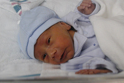
Some degree of ROP appears in more than half of all infants born at 30 weeks pregnancy or younger — a full-term pregnancy is 40 weeks — but only about 5 to 8 percent of cases become severe enough to require treatment. In ROP, blood vessels in the tissue in the back of the eye called the retina begin to grow abnormally, which can lead to scarring and detachment of the retina. Treatment involves destroying the abnormal blood vessels with lasers or freezing them using a technique called cryoablation. Early diagnosis and prompt treatment is the best prevention for vision loss from ROP, which is why the American Academy of Ophthalmology recommends routine screening for all babies who are born at gestational age 30 weeks or younger or who weigh less than 3.3 pounds at birth.

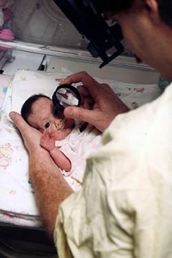
The study evaluated telemedicine for ROP screening during the usual care of 1,257 premature infants who were born, on average, 13 weeks early. About every nine days, each infant underwent screening by an ophthalmologist, who assessed whether referral for treatment was warranted. Those who were referred were designated as having referral-warranted ROP (RW-ROP). Either immediately before or after the exam, a non-physician staff member in the neonatal intensive care unit (NICU) took images of the infant’s retinas and uploaded them to a secure server at the University of Oklahoma, Oklahoma City. Trained non-physician image readers at the University of Pennsylvania, Philadelphia, then downloaded the photos, independently evaluated them following a standard protocol, and reported the presence or absence of RW-ROP.

Through the telemedicine approach, non-physician image readers correctly identified 90 percent of the infants deemed to have RW-ROP based on examination by an ophthalmologist. And they were correct 87 percent of the time when presented with images from infants who lacked RW-ROP. The examining ophthalmologists documented 244 infants with RW-ROP on exam. After referral, 162 infants were treated. Of these, non-physician image readers identified RW-ROP in all but three infants (98 percent). “This is the first large clinical investigation of telemedicine to test the ability of non-physicians to recognize ROP at high risk of causing vision loss,” said Eleanor Schron, Ph.D., group leader of NEI Clinical Applications. “The results suggest that telemedicine could improve detection and treatment of ROP for millions of at-risk babies worldwide who lack immediate in-person access to an ophthalmologist,” she said.

About 450,000 (12 percent) of the 3.9 million babies born each year in the United States are premature. The number of preterm infants who survive has surged in middle income countries in Latin America, Asia, and Eastern Europe. In these parts of the world, rates of childhood blindness from ROP are estimated at 15 to 30 percent — compared to 13 percent in the United States.

retinopathy of prematurity
NICU care providers take photos of a premature baby’s retinas in the NEI-funded e-ROP study of telemedicine for retinopathy of prematurity. Photo credit: Children’s Hospital of Philadelphia

One advantage of telemedicine ROP screening is that it can be done more frequently than screening by an ophthalmologist. “It’s much easier to examine the retina when not dealing with a wiggling baby,” said Dr. Quinn. “If a baby is too fussy or otherwise unavailable when the ophthalmologist visits the NICU, the exam may be delayed until the ophthalmologist returns — sometimes up to a week later.”

Weekly ROP screening — or even more frequently for high-risk babies — is a realistic goal for telemedicine and could help catch all cases needing treatment, according to the report. In the study, imaging was restricted to occasions when an ophthalmologist examined the baby. In practice, hospital staff could implement an imaging schedule based on the baby’s weight, age at birth, and other risk factors. “With telemedicine, NICU staff can take photos at the convenience of the baby,” said Dr. Quinn.

Telemedicine for evaluating ROP offers several other advantages.

Telemedicine may help detect RW-ROP earlier. In the study, about 43 percent of advanced ROP cases were identified by telemedicine before they were detected by an ophthalmologist — on average, about 15 days earlier.

Telemedicine could save babies and their families the hardship and hazards of being unnecessarily transferred to larger nurseries with greater resources and more on-site ophthalmologists. “Telemedicine potentially gives every hospital access to excellent ROP screening,” Dr. Quinn said.

Telemedicine might also bring down the costs of routine ROP screening by reducing the demands on ophthalmologists, whose time is better allocated to babies who need their attention and expertise. In a separate analysis, the study found that non-physicians and physicians had similar success in assessing photos for RW-ROP. Three physicians evaluated image sets from a random sample of 200 babies (100 with RW-ROP based on the eye exam findings; 100 without) using the standard grading protocol. On average, the physicians correctly identified about 86 percent of RW-ROP cases; the non-physicians were correct 91 percent of the time. The physicians correctly identified about 57 percent of babies without RW-ROP; non-physicians were correct 73 percent of the time.

The cost of establishing a telemedicine ROP screening program includes acquisition of a special camera for taking pictures of the retina, training of NICU personnel to take and transmit quality photos, and establishment and maintenance of an image reading center. “As we move along this road, advances in imaging and grading of images may streamline the process even more,” Dr. Quinn said.

The e-ROP Cooperative Group includes the following clinical sites and resource centers:

  • Children’s Hospital of Philadelphia
  • Johns Hopkins University, Baltimore
  • Boston Children’s Hospital
  • Nationwide Children’s Hospital and Ohio State University Hospital, Columbus
  • Duke University (cost-effectiveness center), Durham, North Carolina
  • University of Louisville, Kentucky
  • University of Minnesota, Minneapolis
  • University of Oklahoma (Inoveon ROP Data Center), Oklahoma City
  • University of Texas Health Science Center at San Antonio
  • University of Utah, Salt Lake City
  • Vanderbilt University, Nashville, Tennessee
  • Hospital of the Foothills Medical Center, Calgary, Alberta
  • University of Pennsylvania (data coordinating center and image reading center), Philadelphia

For more information about ROP.

Click to view a video about e-ROP.

NIH logo without bannerNational Institutes of Health
9000 Rockville Pike
Bethesda, Maryland 20892
301-496-4000,
TTY 301-402-9612

Ways to Reduce the Harmful Effects of Sun Glare

During the height of summer sunshine (and heat!), it’s helpful to discuss the importance of eye protection, including ways to reduce the harmful effects of sun glare.

Fundamentally, we need light to see. Approximately 80% of all information we take in is received through the sense of sight. However, too much light – and the wrong kind of light – can create glare, which can affect our ability to take in information, analyze it, and make sense of our surroundings.

Facts about Sunlight

Every type of light has advantages and disadvantages, and sunlight is no exception:

Advantages:

• Sunlight is the best, most natural light for most daily living needs.
• Sunlight is continuous and full-spectrum: the sun’s energy at all wavelengths is equal and it contains all wavelengths of light (explained below).

Disadvantages:

• It is difficult to control the brightness and intensity of sunlight.
• Sunlight can create glare, which can be problematic for many people who have low vision.
• Sunlight is not always consistent or reliable, such as on cloudy or overcast days.

Visible Light and Light Rays

An important factor to consider is the measurement of visible light and light rays, beginning with the definition of a nanometer:

• A nanometer (nm) is the measurement of a wavelength of light.
• A wavelength is the distance between two successive wave crests or troughs:

Wavelength - glare

• A nanometer = 1/1,000,000,000 of a meter, or one-billionth of a meter. It’s very small!

The human visual system is not uniformly sensitive to all light rays. Visible light rays range from 400 nm (shorter, higher-energy wavelengths) ? 700 nm (longer, lower-energy wavelengths).
Visible Light Spectrum - glare
The visible light spectrum occupies just one portion of the electromagnetic spectrum, however:

• Below blue-violet (400 nm and below), is ultraviolet (UV) light.
• Above red (700 nm and above), is infrared (IR) light.
• Neither UV nor IR light is visible to the human eye.

Ultraviolet Light and Blue Light

Ultraviolet (UV) light has several components:

• Ultraviolet A, or UVA (320 nm to 400 nm): UVA rays age us.
• Ultraviolet B, or UVB (290 nm to 320 nm): UVB rays burn us.
• Ultraviolet C, or UVC (100 nm to 290 nm): UVC rays are filtered by the atmosphere before they reach us.

Blue light rays (400 nm to 470 nm) are adjacent to the invisible band of UV light rays:

• There is increasing evidence that blue light is harmful to the eye and can amplify damage to retinal cells.
• You can read more about the effects of blue light at Artificial Lighting and the Blue Light Hazard at Prevent Blindness.

A new study from the National Eye Institute confirms that sunlight can increase the risk of cataracts and establishes a link between ultraviolet (UV) rays and oxidative stress, the harmful chemical reactions that occur when cells consume oxygen and other fuels to produce energy.

Sunlight and Glare

Glare is light that does not help to create a clear image on the retina; instead, it has an adverse effect on visual comfort and clarity. Glare is sunlight that hinders instead of helps. There are two primary types of glare.

Disability glare

• Disability (or veiling) glare is sunlight that interferes with the clarity of a visual image and reduces contrast.
• Sources of disability glare include reflective surfaces (chrome fixtures, computer monitors, highly polished floors) and windows that are not covered with curtains or shades.

Discomfort glare

• Discomfort glare is sunlight that causes headaches and eye pain. It does not interfere with the clarity of a visual image.
• Sources of disability glare include the morning and evening positions of the sun; snow and ice; and large bodies of water, (including swimming pools).

Controlling Glare

You can protect your eyes from harmful sunlight and minimize the effects of glare by using a brimmed hat or visor in combination with absorptive lenses.

• Absorptive lenses are sunglasses that filter out ultraviolet and infrared light, reduce glare, and increase contrast. They are recommended for people who have low vision and are also helpful for people with regular vision.
• Lens colors include yellow, pink, plum, amber, green, gray, and brown. Ultra-dark lenses are not the only choice for sun protection.
• Lens tints in yellow or amber are recommended for controlling blue light.
NoIR Medical Technologies: NoIR (No Infra-Red) filters absorb UVA/UVB radiation and also offer IR light protection.
Solar Shields: Solar Shields absorb UVA/UVB radiation and are available in prescription lenses.
• You can find absorptive lenses at a specialty products store, an “aids and appliances store” at an agency for the visually impaired, or a low vision practice in your area. Before you purchase, it’s always best to try on several different tints and styles to determine what works best for you.

More Recommendations

• Always wear sunglasses outside, and make sure they conform to current UVA/UVB standards.
• Be aware that UV and blue light are still present even when it is cloudy or overcast.
• Make sure that children and older family members are always protected with UVA/UVB-blocking sunglasses and brimmed hats or visors.

Maureen Duffy-editedMaureen A. Duffy, CVRT
Social Media Specialist, visionaware.org
Associate Editor, Journal of Visual Impairment & Blindness
Adjunct Faculty, Salus University/College of Education and Rehabilitation

My New Vision With A Telescope Implant

6/19/14

What it’s like to see with the CentraSight telescope implant

Like many people, I’ve set goals in my life, both professionally and personally and like being an active and engaged member of my community and my country. I love to teach: I taught history, geography and special education for years in Banning, California and now live in Moreno Valley, California, with my wife of 32 years, Kay. I love my country: I’m a proud veteran of both the Army Reserve and the Navy. And I love an open road. My wife and I traveled the country visiting historical monuments in our 32-foot RV. I guess I’ve got what you’d call a real zest for life. But, over the past twenty years, all the things I enjoyed doing in my life, even the simple day-to-day activities, started to decline because I was slowly losing my vision due to age-related macular degeneration. For example, six years ago, my wife took over all the driving because I couldn’t see well enough to drive safely.

Roy Kennedy - telescope implant
Roy Kennedy

That was a real turning point for me. My wife had to help me so much because I just couldn’t see. I needed help shopping because I couldn’t read labels. I started to avoid social situations, like visiting with friends because I was embarrassed that I couldn’t recognize faces any longer. As you can imagine it was heartbreaking for both me and my wife.

But then my doctor told me about a treatment I wasn’t yet aware of called the telescope implant. The device is very small (smaller than a pea!), and it is implanted in one eye to restore vision. My doctor explained that it works like a real telescope in that it magnifies images, which reduces the effect of the blind spot on my straight-ahead vision. The other eye does not get an implant because you need to keep some peripheral vision to help with orientation and balance. This sounded like science fiction! But I wanted to see if it could help me and I decided to give it a try.

I worked with a great team of specialists, who were part of a treatment program called CentraSight. My retina doctor, cornea surgeon, low vision optometrist and a low vision occupational therapist all counseled me about what to expect from the outpatient procedure, particularly afterwards. For example, I learned there was a significant amount of occupational therapy required to adjust and become proficient at using my new vision. I also was warned that my sight would not be like it was in my youth. I wouldn’t be able to do everything I used to nor would I be able to see, differently, the minute I opened my eyes.

I had my surgery in early 2013. The cost for the telescope implant and visits associated with the treatment program were covered by Medicare, which was very helpful. Thinking back, I was nervous on surgery day, but afterwards, I was told by my occupational therapist that I was one of the quickest to recover from surgery. I give lots of credit to my OT folks as well as my wife who helped me with the exercises at home. The most amazing part is being able to see my wife’s face again for the first time in six years! I’ve regained the ability to do many everyday tasks, like reading, working on my computer and watching old Westerns on TV. My wife and I are even back to traveling the open road in our RV (which she drives)!

I would recommend people learn more about the telescope implant to see if it might help them, the way it helped me. There are CentraSight teams across the country. When you call 1-877-99-SIGHT or visit www.CentraSight.com a trained CentraSight information Specialist will point you to the team closest to your home and can even help schedule the appointments for you. The telescope isn’t for anyone, but it can make such a difference in your life. It certainly did in mine.

Roy Kennedy - telescope implantRoy Kennedy

Living With the Argus II

5/27/14

Last year Discovery Eye Foundation spoke to Dean Lloyd, who lost his sight to retinitis pigmentosa (RP), about his experiences with the Argus II “bionic eye.”  After FDA approval, when the article came out, more people across the country have been fitted with the Argus II, using its 60-electrode system to help them regain some part of their vision.  Researchers continue to try and improve upon the “bionic eye” such as this research for a 24-electrode version from Australia.  Here is that article:

Dean Lloyd wearing the ArRgus II
Dean Lloyd wearing the Argus II

As one of only 30 people in the world with a “bionic retina,” Dean Lloyd has gained a bit of notoriety of late. The Argus II Retinal Prosthesis System received US market approval from the FDA on Feb. 14, and Lloyd has been part of the clinical trial since he was implanted with the device in 2007.


Lloyd’s vision difficulties began in the early 1960s, while he was in medical school at the University of South Dakota. He realized he wasn’t seeing the same thing as his classmates when looking through a high-powered microscope. He was misdiagnosed with Usher Syndrome, a rare genetic disorder that can result in deafness, blindness and dementia. While he was later correctly diagnosed with the less-dire x-linked retinitis pigmentosa (RP), he was asked to leave medical school due to his vision impairment.

“I had a moment of self-pity, then I needed to do some introspection to figure out how to find my future,” he says. “I decided I can’t give up on life. My brain works well … If your brain works, it can solve a lot of difficult problems. I realized I would survive.”

Seeing Stars

Armed with a BS in chemistry and an MS in bio-chemistry, Lloyd became a research chemist, then a software engineer. At the time, RP was having only one real effect on his life: reduced night vision. “I always thought people were seeing more stars than I saw. They’d see the big dipper and little dipper, and I didn’t see any dippers,” he recalls. Other than being extra-careful when driving at night, Lloyd’s daily life remained relatively unaffected until 1974, when he developed cataracts at age 34.

Lloyd’s wife decided she did not want to deal with any impact the disability would have on his ability to support their family. “She left and got the best divorce lawyers,” he says. “The court thought, because I was visually impaired, I wouldn’t be able to take care of my children. There were a lot of issues around disability and parent-ability in the mid- to late 70s.” Because of his vision loss, Lloyd lost custody of his two children. Believing disabled people were not treated fairly and seeing “the power of the court over people in their everyday lives,” Lloyd decided to go to law school. He passed the bar in 1982, “as a disabled person in a closed room,” and has been practicing law for more than 30 years.

He underwent two cataract surgeries, and a pair of convex lenses he wore on his nose was used in place of the actual lenses that had been removed from his eye. He had no difficulty with daytime mobility for nearly 14 years. But in 1989, he lost image formation. “I got an edema within the macula area, which didn’t go away,” he says. “I lost the ability to form images — that’s the point when you lose the opportunity for safe mobility.”

Lloyd had already been practicing law for seven years, and RP didn’t stop him. “I had quite a bit of experience in the courtroom,” he says. “I am fortunate to have a good memory, and I memorized everything I used there. I usually work with a junior or associate attorney; I do all the talking, and they handle all the documents and things. It didn’t change my career.”

A cane helped with his mobility, and for a few years, he says, “A doctor at Stanford pulled me in for some non-FDA experiments, because he knew I was a four-eyed guinea pig and was willing to try anything that might help RP.” None worked.

Lloyd was involved with several vision-related organizations and followed the progression of research and experiments to see if any might benefit him. In early 2007, he attended a Foundation Fighting Blindness meeting run by his daughter, who has “the recessive expression for RP.” The speaker that day was Dr. Jacque Duncan of UCSF, who interviewed all the attendees. Soon after, she contacted Lloyd to ask him to participate in the Argus II clinical trial.

He agreed immediately: “I figured if I lost the eye, I wouldn’t lose much, because it didn’t work anyway.”

The Bionic Retina

The Argus II Retinal Prosthesis System consists of three parts: an implant, glasses and a belt-worn video-processing unit (VPU).

The implant is surgically implanted in and on the eye. It includes an antenna, an electronics case and an electrode array. The glasses contain a tiny video camera that captures a scene. The video is sent to the VPU, where it is processed and turned into electrical stimulations — instructions — that are sent back to the glasses through a cable. The glasses then transmit the instructions wirelessly to the implant antenna, which sends signals to the electrode array, which emits electric pulses. These pulses bypass damaged photoreceptors (RP damages photoreceptors and impedes image formation) and stimulate the retina’s remaining healthy cells, which transmit the information along the optic nerve to the brain. If all goes well, it creates the perception of patterns of light, which wearers can learn to interpret as visual patterns.

Chicken Dance

Duncan, working with another UCSF doctor, had Dr. Eugene de Juan put the Argus II implant in Lloyd’s right eye on July 17, 2007. A few weeks after an initial infection subsided, he did his first test walk with the device outside, looking for boundaries and points of light.

“Human eyes naturally move around, using a rapid ‘saccade’ movement,” Lloyd explains. “With the device, you have to physically move your head back and forth to find boundaries — like a chicken, which does not have moving eyes. They move their heads back and forth to see things. During my walk, the field-service worker would say, ‘Dean, Dean, more chicken movement.’”

After about a year and a half spending a half-day a week at UCSF for the trial, he had a second operation. “The device didn’t satisfy me,” he says. “I went through all that, and all I could see was a little point of light. Only nine of 60 electrodes were working. I said, ‘This isn’t going to cut it for me, because I want to see images.’ They reset the implant closer to the retinal tissue, and that dramatically improved the device.”

Lloyd conducted his own tests of the device at home. “I was one of first to use it to sort socks. I wear black socks to court and white socks to the gym. I went home after the implant and wanted to see how I could make it practically useful. So I went to my socks and found the device worked. I always got the white ones right!”

While he is generally pleased with the Argus II, the most he will say is, “It shows potential.”

“The improvement was not very good at first. I wasn’t very impressed. There was a rumor going around that the implant restored vision. My vision wasn’t restored at all; I was not ascertaining images,” he says. “I do see boundaries. I can create an image. I know what a car should look like. I know what a tree should look like. I know what houses should look like. I know what objects should look like, and I have those images as memories in my brain. With this device, you can start to create an image by going back and forth, checking the boundaries and borders. We don’t have natural saccade movement helping us. The camera is right above my nose in a fixed position. It doesn’t move. You can create an image, but it takes a lot of time and a lot of work. It’s a labor-intensive task, and you have to have a good memory.”

The Argus II was approved by the FDA in February, and Lloyd is actively involved with Second Sight, the company that makes the device: “I am helping with new improvements,” he says. “I want this device to work. I didn’t go through all this for nothing.” He hopes an improved version will be approved this coming September, and he doesn’t rule out getting a new implant in the future if the improvements are worth it. To be honest, he says, “I find it annoying to move like a chicken.”

Susan DeRemerSusan DeRemer, CFRE
Vice President of Development
Discovery Eye Foundation

The Evolving Contact Lens

4/22/14

Contact lenses give a person the ability to see without glasses. If you have keratoconus, they are essential for seeing as regular glasses don’t work with an irregularly shaped cornea. But lately these relatively simple lenses have created a whole new world where they can dispense eye medication, measure blood glucose levels and even help the blind see.

Courtesy Google
Courtesy Google

Monitoring Blood Sugar
You have heard about Google Glasses, but Google is looking beyond the smartphones of eye wear to monitoring health. They are currently working on a lens with tiny wireless chips and glucose sensors that are sandwiched between two lenses. They would monitor glucose levels once a second and use tiny LED lights, also inside the lenses, to flash when the levels are too high or low. And how big are these electronics? They are no larger than a speck of glitter, with a wireless antenna that is thinner than a human hair. While they are still in development – Google has run clinical research studies and is in discussions with the FDA – it could make blood sugar monitor far less invasive than pricking your finger several times a day.

Drug Delivery for Glaucoma
Getting glaucoma patients to regularly use their eye drops to regulate the pressure in their eyes has always been a problem. They forget, don’t want to be bothered, or have a hard time getting the drops into their eyes. This could change with two research projects exploring the use of contact lenses to deliver medication over a prolonged period of time.

Researchers at Massachusetts Eye and Ear/Harvard Medical School Department of Ophthalmology, Boston Children’s Hospital, and the Massachusetts Institute of Technology who are working on a lens designed with a clear central area and a drug-polymer film made with the glaucoma drug latanoprost, around the edge to control the drug release. These lenses can be made with no refractive power or the ability to correct the refractive error in nearsighted or farsighted eyes.

Another team from University of California, Los Angeles have combined glaucoma medication timolol maleate with nanodiamonds and embedded them into contact lenses. When the drugs interact with the patient’s tears, the drugs are released into the eye. While the nanodiamonds strengthen the lens, there is no difference in water content so they would be comfortable to wear and allow oxygen levels to reach the eye.

Seeing in the Dark
Researchers out of the University of Michigan have developed an infrared sensor that could eventually be used in the production of night vision contact lenses. Thanks to graphene, a tightly-packed layer of carbon atoms, scientists were able to create a super-thin sensor that can be stacked on a contact lens or integrated with a cell phone.

Stem Cells for Cornea Damage
Researchers in Australia are working on a way to treat corneal damage with stem cell infused contact lenses. Stem cells were taken from the subject’s good eye and then plated them onto contact lenses (if there is a defect in both eyes, stem cells are taken from a different part of the eye). After wearing for about two weeks the subjects reported a significant increase in sight.
Braille-Tracile-Contacts
Helping the Blind See
And what good are contact lenses if you are blind? At Bar Ilan University in Israel researchers are creating special lenses that translate images into sensations felt on the eye. It works by taking an image with a smartphone or camera, it is then processed and sent to the contact lens. The custom-made lens is fitted with a series of electrodes that use small electric impulses to relay shapes onto the cornea, similar to braille. After some practice, test subjects were able to identify specific objects.

In expanding the uses of contact lenses, these projects seem to be just the beginning, all reported in the first four months of this year. Researchers and developers are working together to find more and better ways help with vision and medical issues, using contact lenses.

Susan DeRemerSusan DeRemer, CFRE
Vice President of Development
Discovery Eye Foundation

Wavefront Sensing Applied to Custom Contact Lens Research in Keratoconus

4/10/14

During a trip to the optometrist or ophthalmologist, a patient will encounter the process of subjective refraction.  This technique involves the clinician asking the patient to make a series of judgments (which is better, one or two?) about the clarity of their vision when looking through a series of lenses.  The choices that the patient makes guide the clinician in identifying an optical prescription which is typically made up of sphere, and potentially, cylinder lenses.

Why is it that glasses don’t always work for patients with keratoconus?

In many instances, individuals with keratoconus do not achieve excellent visual performance with spectacles or traditional soft contact lenses.  One cause for the failure of these corrections is that the changes in corneal shape that accompany keratoconus induce refractive errors which traditional spectacles simply cannot correct.  So, even when sphere and cylinder in the keratoconic eye are well-corrected, these “other refractive errors” or “other aberrations” remain uncorrected and can lead to a blurred retinal image and blurred vision.  Collectively these other aberrations can be referred to as higher order aberration, while the aberrations that are typically corrected with spectacles and soft contact lenses are referred to as lower order aberration.

What kinds of higher order aberrations are present in keratoconus:

Pantanelli et al. have stated that the level of higher order aberration present in an eye with keratoconus is, on average, approximately 5.5 times higher than the level experienced in a control group.  In an effort to visualize higher-order aberration data, they are commonly represented graphically as shown in the figures below.  Examples of higher order aberration measured in one normal eye are shown in figure A, while an example of higher order aberration from one keratoconic eye are shown in figure B.  The circular nature of the map denotes the boundary of the measurement, which is defined by the round pupil of the eye.  A majority of the higher order aberration map in figure A is green (denoting a relative absence of higher order aberration).  However, the map in figure B displays a much larger variation in color, indicating the presence of higher order aberration  in this individual keratoconic eye in a greater quantity than the normal eye shown in figure A.

Figure A - normal-keratoconus
Figure A – normal
Figure B - keratoconus
Figure B – keratoconus

A wavefront aberration map of the “other aberrations” or higher order aberrations of two eyes. Figure A is an example of data for a normal eye and figure B is an example of data for an eye with keratoconus.

If refraction is not capable of quantifying higher order aberrations, how are they measured?

One method for obtaining the information regarding higher order aberration shown above is with a wavefront sensor.  The wavefront sensor objectively (without patient feedback) collects information on the optical performance of the eye that can be used to calculate the amount of both lower and higher order aberration present.

Laboratory-based research related to custom contact lenses:

Several investigators in the laboratory (e.g. Katsoulos et al., Sabesan et al., Chen et al., Marsack et al.) have reported on work that attempts to further reduce higher order aberration by targeting the eye-specific higher order aberration seen in a given keratoconic eye.  The general philosophy behind these customized lenses is that the aberration pattern measured with the wavefront sensor is a more complete optical prescription for implementation of a custom contact lens.  Figure C demonstrates, in principle, the optical properties of a contact lens designed to correct the higher-order aberration in figure B.  Where the map of the eye (figure B) is red, the map of the correction (figure C) is blue, and vice versa.  When the lens is worn, the net effect as light propagates through the lens-eye system is the cancellation of the higher order aberration in a targeted manner.

Figure C -keratoconus correction

In principle, this figure pictorially represents the higher order optical properties of a contact lens designed to fully correct the higher-order aberration of the eye represented in figure B.

What is next:

Investigators continue to push the technology behind custom contact lenses for keratoconus towards clinical relevance.  However, like every novel intervention strategy, we must manage our expectations.  Complexity in measuring keratoconic eyes, a need for specialized equipment and expertise to design and manufacture the lenses, the infrastructure needed to coordinate the clinical exam and manufacture efforts and cost associated with the process are a subset of the barriers that must be removed if this type of correction is to become more mainstream.  For this reason, it is my opinion that if/when these corrections become commonly available in the clinic, they will likely add to, and not replace, existing forms of corrections that patients and clinicians now utilize to correct vision.

jmarsack-bio-picJason Marsack, PhD
Research Assistant Professor
University of Houston, College of Optometry.
Dr. Marsack’s work focuses on the relationship between visual performance
and optical aberration in individuals with highly aberrated eyes.

End of the Day Syndrome

4/2/14

“Dr. S., my eyes are red and burning at the end of my work day.”

“Patient, what sort of work do you do?  Tell me something about your work conditions.”

“I am a computer graphics artist.  I sit and stare at my twenty-seven inch HD screen for hours on end gently adjusting the composition of each pixel.  My studio is air-conditioned but not humidified, so after some hours of work, I feel dry as a bone.”

“One more question…can you cry tears?  Say, when you peel and slice an onion?”

Rule of 20 - blinking

The need to blink

Blinking is a complex function of the eyelids that when completed results in a clean, refreshed, re-wetted corneal surface.  The tears that are washed across the outside of the eye with each blink bring oxygen and other nutrients to the outer cell layer aiding in the rebuilding and revitalizing of the surface tissue.

Blinking is characterized by a full sweep of the upper lid over the eye to meet the lower lid.  The completion of this motion is performed gently without squeezing.  And, to be effective full eye closure needs to be repeated fairly often.  Blink rates vary according to investigators but most sources report an average of between six and ten full blinks per minute under normal viewing circumstances.

The anti-blink problem of our generation

In olden times – say the years between 1750-1950 – the most aggravating problem to the ocular surface was a good book or intense study.  The reader would concern himself with the text at hand and slowly his eyes would dry until a “rest break” was necessary.

Environmental or vocational changes to our lifestyle over the generations have promoted reduced blink rates.  Most recently in this negatively developmental progression is the effect of the television screen, the CRT, the LED screen, the handheld and pocket computer on the blink rate.  It appears that as attention level increases, blinking suffers.  First the eyes close less, then incompletely, and finally rarely only when surface dryness drives the individual to desperate measures.  He must blink or (so he feels) his eyes will pop out of their sockets.

Adding insult to injury increasingly over the decades is air conditioning – both heating and cooling – when not humidified.  Staring at console screens in dry environs speeds the desiccation of the cornea and results in discomfort.

The surface of the eye is a biological system.  Living systems require some degree of moisture.  If the cells of the eye – or any biological surface — are permitted to dry out, they will die.  Dead corneal cells fall off the cornea and float in the tears on the surface of the eye until washed away with a blink.  Until the surface is cleaned the dead cells are considered by the eye to be foreign bodies with the consequent irritation and induced reflex to blink.

When cells die and fall off, the underlying nerve endings send pain signals to the nervous system.  The sensation can be felt as pain, burning, or mere irritation or itching depending upon the severity of cell loss.

How to handle environmentally induced dry eye

After the ocular surface is dry most treatments will seem to make matters worse:  to cause burning and stinging, perhaps, even more than the dry eye itself.  Any tear substitute, any amount of blinking will be irritating at first. But, that is really all that can be done at this stage:  wetting and blinking.

Prevention

As in many conditions, the best treatment, in fact a cure, for recurrent environmentally induced dry eye is prevention.  For the eye that has a naturally flowing tear supply, the act of blinking is the surest prevention to stinging and burning after a day’s work at the computer.  Additionally, many sources recommend using the ‘rule of 20’:  after each twenty minutes of work, look up from the text or away from the screen; blink and refocus on the page twenty times.  This repetitive exercise simultaneously re-wets the eye and relaxes the focusing mechanism of the eye.

The result is relaxed and comfortable eyes that can continue to provide important and high quality information for longer hours of work.

Bezalel-SchendowichBezalel Schendowich, OD
Chairperson and Education Coordinator, JOS
Fellow, IACLE
Member, Medical Advisory Board NKCF
Sha’are Zedek Medical Center, Jerusalem, ISRAEL

Help for Computer Users

Working long hours in front of the computer requires a fairly unchanging body, head and eye position which can cause discomfort.  Correct working position, periodic stretch breaks, frequent eye blinking, artificial tears for lubrication are all very important.  However, it’s not always easy to remember this when you are engrossed in work. Here are a few fun, free and easy-to-install “break reminders” to help:

WorkSafe Sam - break reminder
WorkSafe Sam
WorkSafe Sam is a desktop tool that provides stretching tips to help reduce eye and muscle strain for office workers (clicking on this link will open a file on your computer because this is a zip file).

Workrave is break reminder program that alerts you to take “micro-pauses” and stretch breaks.

Take Your Break is another break reminder designed to prevent or minimize repetitive strain injury, computer eye strain and other computer related health problems.  It has a friendly interface and a tray icon status indicator.  It runs quietly in the background, monitoring your activity and reminding you to take regular breaks.

And remember to blink.  Blinking cleans the ocular surface of debris and flushes fresh tears over the ocular surface. Each blink brings nutrients to the eye surface structures keeping them healthy. The flow of tears is responsible for wetting the lower third of the cornea. This is very important in KC, since this area is generally below the bulge of the cone and in many cases irritated by wobbly RGP lenses.  Maybe your job requires hours of work at a computer. Maybe you like to spend your free time surfing the internet. Whatever the reason, your body is probably feeling the effects of spending too much time staring at a computer monitor, which could result in Computer Vision Syndrome (CVS).  The most common symptoms are: eye strain, dry or irritated eyes,redness in eyes,difficulty in refocusing eye,neck pain,double vision,blurred vision, fatigue, and headaches.

Please join us on Thursday when Dr. Bezalel Schendowich will be providing a detailed insight into the importance of blinking, going beyond computer usage.

CathyW headshotCathy Warren, RN
Executive Director
National Keratoconus Foundation

Implantable Miniature Telescope Update

The Macular Degeneration Partnership, a program of the Discovery Eye Foundation,  has received numerous questions about the implantable miniature telescope (IMT) since it was approved by the FDA in 2010.

Implantable Miniature Telescope
The IMT is becoming more widely available now. The IMT is a tiny telescope implanted inside the eye that may benefit older adults with advanced AMD. Smaller than a pea, this device is proven to restore sight and quality of life in eligible candidates. Unfortunately, the inclusion criteria to be eligible for the device are narrow.  Most importantly, the IMT can only be implanted into an eye that has not had a cataract removed yet.   We encourage you to review the below information to see if you or a loved one might be a candidate for this procedure. Approximately 2 million Americans have advanced forms of AMD, which is the leading cause of blindness in people over the age of 65. When an individual has severe wet macular degeneration, or dry AMD with geographic atrophy, it is sometimes called “end-stage AMD”. Patients with end-stage AMD have a central blind spot or missing area in their vision. But, despite the availability of drug treatments that slow the progression of AMD, the number of people with end-stage AMD is expected to double by the year 2050.

Specifically, the telescope implant uses micro-optical technology to magnify images which would normally be seen in your “straight ahead,” or central, vision. The images are projected onto the healthy portion of the retina not affected by the disease, making it possible for patients to see straight ahead. The procedure is performed on one eye only, and involves removing the eye’s natural lens and replacing it with the tiny telescope implant. This is similar to the surgery performed to remove a cataract, which is a clouding of the natural lens. The other eye remains as is to preserve peripheral vision, which is important for balance and orientation. The surgery is done in an outpatient setting by a specially-trained ophthalmologist called a cornea/cataract surgeon. The telescope implant is FDA approved and available through Medicare.

Although the telescope implant is not a cure for AMD, studies showed that in general patients were able to see 3 to 4 lines better on the eye test chart and demonstrated improved quality of life on the National Eye Institute Visual Functioning Questionnaire. Two multi-year clinical studies enrolled over 225 patients to evaluate the safety and efficacy of the telescope implant used in the CentraSight treatment program.   To be considered a candidate for the telescope implant, an ophthalmologist must first confirm that you:

• Have irreversible, End-Stage AMD resulting from either dry or wet AMD
• Are no longer a candidate for drug treatment of your AMD
• Have not had cataract surgery in the eye in which the telescope will be implanted
• Meet age, vision, and cornea health requirements

Some people with end-stage AMD may not be a candidate for a telescope implant. Patients and their physicians will assess if the benefits of the procedure outweigh the potential risks to decide if this treatment option is right for them.

CentraSight is the program that guides people with end-stage AMD through the telescope implant evaluation, surgery and rehabilitation process.  While the out-patient procedure is quick, patients also must commit to a comprehensive occupational therapy program to learn how to use their new vision (and way of seeing) in daily life. Click here to watch a video that shows how the implantable telescope works.

“After surgery, one of the most important aspects of the telescope implant procedure is the rehabilitation,” said Dr. Marjan Farid, Associate Clinical Professor of Ophthalmology at the University of California-Irvine School of Medicine. “Specially trained optometrists and occupational therapists work with patients to teach them how to use their new vision because there are different techniques involved when you are sitting still (for example, reading or watching TV) than when you are moving around, such as walking or cooking.”

The CentraSight treatment program is coordinated by retina specialists who treat macular degeneration and other back-of-the-eye disorders.  Before deciding to have the surgery, a special vision test is given in the office.  A device simulates what a person may expect to see once the telescope is implanted to determine if the potential improvement will meet the patient’s expectations. Once the telescope has been implanted by an eye surgeon, the patient will need to work with vision rehabilitation specialists (approximately 6 to 12 weeks) to learn how to use their new vision in their everyday activities. Risks include all those associated with cataract surgery, such as postoperative inflammation, raised intraocular pressure, corneal swelling, and the potential for comprised corneal health.

“The first patient whom I implanted with this telescope over a year and a half ago states that she can now recognize the faces of her children and grandchildren,” said Dr. Farid. “For patients with AMD, face recognition of loved ones is a major improvement in the overall quality of their life.”

CentraSight treatment centers are available across the nation. Patients can call 1-877-99-SIGHT to find one in their area.

Judi Delgado headshotJudith Delgado
Executive Director
Macular Degeneration Partnership