A New Commitment to Vision

Meet Tom Sullivan – DEF’s Ambassador of Vision

Over the last 40 years, I’ve been committed to working on behalf of blind children and their families.  My involvement has spanned the entire gamut of participation – from direct involvement in the classroom and counseling parents to hosting organized 10K races and celebrity golf tournaments that carried my name.  In that time, my wife and I raised just over $8 million thanks to the generosity of so many people.  Though my commitment to this cause has not changed, I’ve chosen to take on a new challenge that has in every way re-energized my passion. 

I’ve recently become the Ambassador of Vision for the Discovery Eye Foundation (DEF), a remarkable organization that funds cutting edge research that I believe someday will eliminate many forms of blindness.  The principle reason for my enthusiastic commitment is largely due to the fact that DEF directly funds researchers and avoids institutional restrictions.

As of this blog, DEF is engaged in ongoing efforts to understand 5 main eye diseases – Retinitis Pigmentosa, Macular Degeneration, Keratoconus, Diabetic Retinopathy, and Glaucoma.  I can honestly tell you that breakthroughs are not only on the horizon, but in many cases they are imminent.  Over the weeks and months I’ll be telling you much more, specifically about our individual research projects.  

Any help you may choose to give on behalf of people struggling with vision loss will be deeply appreciated. I look forward to having many of you join my fight for sight. Your help can make a difference! Click here to donate.


Tom Sullivan
DEF’s Ambassador of Vision
sullivanvision.com


Who’s On First For My Eye’s

The Difference Between an Optometrist and an Ophthalmologist

There are a number of players on your eye-health team, among them your optometrist and your ophthalmologist. It’s not always obvious, however, what position each plays and when you should call on which professional. 


Optometrists 

An optometrist holds a doctor of optometry (OD) degree. An optometrist generally completes a four-year college program in the sciences, plus four years of postgraduate professional training in optometry school.

Optometrists examine eyes for vision and health problems, and correct refractive errors with glasses and contact lenses. Some optometrists also provide low vision care and vision therapy. In the United States, optometrists may prescribe medications to treat certain eye problems and diseases, and their scope of care can differ by state. While not licensed to perform eye surgery, optometrists can play a role in pre- and post-operative care, if you have eye surgery performed by an ophthalmologist.


Ophthalmologists

An ophthalmologist is a medical doctor (MD) or a doctor of osteopathic medicine (DO), who specializes in eye and vision care. Ophthalmologists complete four years of college, four years of medical school, one year of internship and at least three years of residency in ophthalmology. Some ophthalmologists also have one or two years of additional training to become specialists in cornea, retina, glaucoma, oculoplastics, pediatrics or neuro-ophthalmology.

Ophthalmologists perform eye exams, diagnose and treat diseases, prescribe medications and perform eye surgery. They also prescribe corrective lenses.


Which one do I call?

Both optometrists and ophthalmologists perform routine eye exams, and are both trained to detect, diagnose and manage eye diseases. Both can prescribe corrective lenses, and both must fulfill continuing education requirements to maintain their licenses.

If your eyes are healthy, which type of professional to see is a matter of personal preference. If you already have a medical eye problem, such as diabetes, or a family history of AMD or glaucoma, you should consider seeing an ophthalmologist. While most optometrists offer medical treatment for common eye problems and chronic eye diseases, such as pink eye, dry eye or allergies, certain eye disorders require treatment by an ophthalmologist, particularly if you need surgery, laser therapy or other specialized care.

In some cases, your optometrist and ophthalmologist may work as a team. In such a setup, the optometrist is the equivalent of your primary-care doctor, who monitors your ongoing condition and care, and the ophthalmologist is your specialist, who manages your condition medically and/or performs surgery. For routine care, you may want to check with your insurance provider to see which, if either, professional they cover and for what services.

World KC Day

World KC Day is November 10th! 

World KC Day is an awareness day to help bring a spotlight to keratoconus and honor those that live and cope with keratoconus every day.

Keratoconus, often abbreviated as “KC”, is a non-inflammatory eye condition in which the normally round dome-shaped cornea progressively thins causing a cone-like bulge to develop. This results in significant visual impairment.

Do you or someone you know have KC?

Here are some ways you can join the National Keratoconus Foundation and thousands around the world in spreading the word about keratoconus:

⇒ Join the NKCF Twibbon campaign
⇒ Add yourself to the World KC Map
⇒ Post about keratoconus and use #worldkcday as your hashtag
⇒ For more information about World KC Day visit worldkcday.com

The National Keratoconus Foundation is a program of the
Gavin Herbert Eye Institute at the University of California, Irvine.

View Video

What Is Happening In the Gas-Permeable Contact Lens Industry

The Gas-Permeable Contact Lens

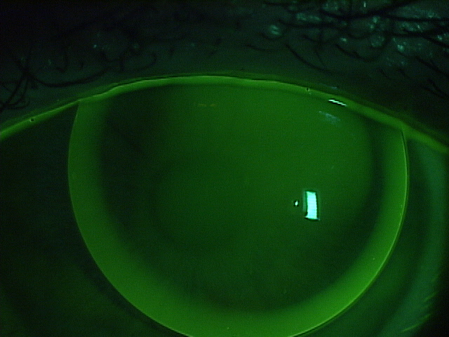
The mainstay of treatment for our patients with keratoconus are gas-permeable lenses.  Corneal gas-permeable (GP) lenses have been the treatment of choice for over 40 years and fit approximately two-thirds the size of the cornea.  Corneal GP lenses translate and pump tears and oxygen under the lens with each blink.  Hybrid lenses have a GP center bonded to a soft skirt that cushions and centers the lens.  Scleral lenses are very large diameter lenses that completely vault the cornea and land on the relatively insensitive scleral tissue.  Whether they are corneal GP, hybrid, or scleral lenses, the commonality is that all of these strategies use the optics and rigidity of GP materials to provide the best vision and comfort for this challenging condition.

gas-permeable contact lens
Well-fit corneal GP lens with sodium fluorescein dye

The way that the GP industry works is that GP button manufacturers (there are 6 in the US) sell the raw material, or button, to independent laboratories (there are 39 in the US) who then craft the button using industrial lathes into hundreds of lens designs using their own intellectual property.   GP buttons are used to make corneal GPs, hybrid, and scleral lenses.  Doctors who are skilled in fitting and evaluating the lens designs craft custom made GP lenses for their patients for whatever purpose benefits the patient.

Not many people know that the GP lens industry is on fire right now.  The turmoil began when Valeant Pharmaceuticals purchased Boston Products.  Boston Products manufactures the raw material of GP lenses, GP buttons, and held around 80% of the US market share.

The independent laboratories have a trade association called the Contact Lens Manufacturer’s Association (CLMA) who have an educational wing called the Gas-Permeable Lens Institute (GPLI).  The GPLI is universally beloved by practitioners because its primary function is to educate doctors to become better doctors in a non-branded, good-of-the-industry format.  No lens design is favored over any other.  Education is free to all doctors and expertise in specialty lenses is stressed, so that patients are placed in skilled hands.  Jan Svochak, president of the CLMA, says, “The CLMA represents a longstanding group of Independent Contact Lens Manufacturers working collaboratively where we have shared goals. These include educational resources through the GPLI that work closely with Eye Care Practitioners and Educational Institutions as well as a dedication to protecting and advancing utilization of custom manufactured contact lenses.”

Next Valeant dropped the hammer on the industry.  Overnight and for no apparent reason, they sent a letter to all of the independent laboratories announcing that they were increasing the price on scleral lens-sized Boston buttons by an astounding 60% (and other buttons by multiples).  Simultaneously, they announced that they were dropping out of the CLMA.  This move stunned the CLMA member labs as it blocked them from supplying Paragon CRT lenses to doctors.  Being the market leading GP button manufacturer, dropping out of the CLMA essentially defunded the GPLI and ensured the immanent collapse of the CLMA.

The price increase sent shock waves throughout the industry.  There was a simultaneous but independent reaction from many of the key-opinion leading optometrists who fit GP lenses.  The problem with any increase in price on the GP button level is that these price increases are passed down the line through the laboratories, the doctors and eventually, to the patients.  Valeant saw a huge backlash from optometrists who essentially stopped prescribing their materials.  They admitted making a mistake, and lowered the cost of the buttons, but interestingly, not to the original level.  Instead, there was an average 16% increase in the cost of scleral lens buttons to the laboratories.  Similarly, Valeant did not rejoin the CLMA.  The cost increase has been reported to fund Valeant brand specific education.

The other members of the CLMA came together and saved the association and the GPLI.  Additionally, a key competitor to Valeant, Contamac, rejoined the CLMA.  Contamac is a button manufacturer who formerly held around 8% market share of GP buttons.  At present, key sources within the industry believe that the market share has essentially flip-flopped, so that now, Contamac has rapidly gained market share of the GP button space as doctors have largely abandoned Boston materials in protest of these moves.

In a reactionary panic, Valeant has most recently written to the CLMA, asking to rejoin, but paradoxically with demands.  The CLMA is currently reviewing whether to allow Valeant to rejoin and under what terms.  Long term, it is beneficial for the industry for everyone to work together for the common good. It is unfortunate that a large company has come into the keratoconus treatment area and is raising prices without providing any real value, such as research and development into newer and better tools.  Companies like Valeant ultimately need to realize that they are not in control of an industry.  The patients and doctors are.


Dr. Sonsino is a partner in a high-end specialty contact lens and anterior segment practice in Nashville, Tennessee.  For over 12 years, he was on the faculty at Vanderbilt University Medical Center’s Eye Institute.  Dr. Sonsino is a Diplomate in the Cornea, Contact Lens, and Refractive Therapies Section of the American Academy of Optometry (AAO), chair-elect of the Cornea and Contact Lens Section of the American Optometric Association (AOA), a fellow of the Scleral Lens Education Society, board certified by the American Board of Optometry (ABO), and an advisory board member of the Gas Permeable Lens Institute (GPLI).  He lectures internationally, publishes in peer-review and non-peer-reviewed publications, and operates the website: TheKeratoconusCenter.org.  He consults for Alcon, Art Optical, Allergan, Johnson & Johnson, Optovue, Synergeyes, Visionary Optics, Visioneering, and formerly for Bausch & Lomb.

3/30/16

Sonsino Headshot

Jeffrey Sonsino, OD, FAAO
The Contact Lens Center at Optique Diplomate
Cornea, Contact Lens, and Refractive Therapies,

Having Trouble with Your Scleral Lenses?

Scleral lens have become very popular and while many enjoy the comfort and vision correction they provide, some find it difficult to apply (insert) and remove these large diameter RGP lenses. If you are one of the many who are having difficulty managing to get your scleral lenses in or out, there are a number of tools available to help.

The Scleral Lens Education Society website provides a wealth of information about the care and handling of these lenses trouble with your Scleral Lenses as well as an excellent video. There are photos demonstrating various ways to hold the lens while applying it …some you may not have tried! If you have trouble with your schleral lenses, see below.

Troubleshooting tips and tricks:

    • If you are unable to maintain fluid in the bowl of the lens as you bring it towards your eye, make sure that your face is fully parallel to the floor. It may seem like you are nearly standing on your head when you’re in the correct position to apply the lens.
    • Lid control is essential; use one hand to hold lids completely out of the way, and don’t release the lids until the lens is actually fully in place and the plunger (or your finger tripod) has been removed.
    • If you are unable to successfully apply a solid lens with saline, you could practice applying the lens after filling the bowl of the lens with Celluvisc™ or another non-preserved viscous lubricant. These viscous lubricants will blur your vision compared to saline, however, so you may simply want to use them to practice lens application. Once you’ve mastered this step, switch to saline to give you better vision.
    • Try to keep both eyes open as you apply your lenses. This may also help you to position the lenses correctly.
    • If you are using a bulbed (DVM) plunger, and can see the opening in the center of the suction cup, look directly at the hole as you bring the lens into position. This will help you to position the lens correctly.

    Many find the DVM plungers helpful. They are readily available at your doctor’s office and online. These are just a few of the places to find them: DMV Corp, Dry Eye Zone, and Amazon.

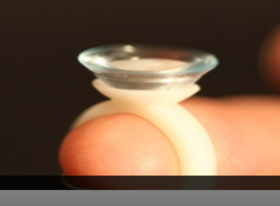
    trouble with your schleral lensesAnother variation to the standard lens inserters is a ring-style lens applicator by EZI Scleral Lens. It was designed by a post-transplant patient who like so many, had trouble inserting his scleral lens without getting a bubble. Read Tim’s story.

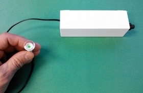
    If you have tried the above techniques and still have trouble applying scleral lenses there are a number of devices available that may help. Dalsey Adaptives has developed the See-Green devicetrouble with your schleral lenses that can be used to help successfully apply scleral contact lenses. The See-Green system comes with a stand that holds a lighted plunger (Figure 2). Using this system, you don’t hold the lens, you lower your eye onto it, which leaves both hands free for improved lid control. The light at the center of the plunger is used as a target to help you position the lens centrally on the eye. Click here to see the detailed instruction sheet.

    Scleral lenses offer good vision and comfort but can be challenging to manage. Discuss these options with your eye care professional to get his or her recommendation for your specific situation.

    1/28/16


    CathyW headshotCathy Warren, RN
    Executive Director
    National Keratoconus Foundation

Top 10 Articles of 2015

eye facts and eye disease
In looking at the many articles we shared with you in 2015, we found that your interests were varied. From the science of vision, eye facts and eye disease to helpful suggestions to help your vision.

Here is the list of the top 10 articles you read last year. Do you have a favorite that is not on the list? Share it in the comments section below.

    1. Rods and Cones Give Us Color, Detail and Night Vision
    2. 20 Facts About the Amazing Eye
    3. Understanding and Treating Corneal Scratches and Abrasions
    4. 32 Facts About Animal Eyes
    5. 20 Facts About Eye Color and Blinking
    6. When You See Things That Aren’t There
    7. Posterior Vitreous Detachment
    8. Can Keratoconus Progression Be Predicted?
    9. Winter Weather and Your Eyes
    10. Coffee and Glaucoma: “1-2 cups of coffee is probably fine, but…”

Do you have any topics you would like to see discussed in the blog? Please leave any suggestions you might have in the comments below.

1/7/16


Susan DeRemerSusan DeRemer, CFRE
Vice President of Development
Discovery Eye Foundation

The Importance of An Eye Exam

Why You Need An Eye Exam

The end of the year is fast approaching – when was the last time you had an eye exam? Was it a comprehensive eye exam?
eye exam
To keep your eyes healthy and maintain your vision, the American Optometric Association (AOA) recommends a comprehensive eye exam every two years for adults ages 18 to 60, and annual exams for people age 61 and older. However, if you have a family history of eye disease (glaucoma, macular degeneration, etc.), diabetes or high blood pressure, or have had an eye injury or surgery, you should have a comprehensive exam every year, unless otherwise indicated by your doctor.
Also, adults who wear contact lenses should have annual eye exams.

An important part of the comprehensive eye exam is the dilated eye exam to look inside your eye. Drops are placed in each eye to widen the pupil and allow more light to enter the eye. This gives your doctor a clear view of important tissues at the back of the eye, including the retina, the macula, and the optic nerve. This allows for early diagnosis of sight-threatening eye diseases like age-related macular degeneration, diabetic retinopathy, glaucoma, etc.

To better understand the importance of the dilated eye exam, here is a video from the National Eye Institute (NE) that explains what to expect.

At the end of your comprehensive eye exam your doctor should raise any concerns he has with you. But it is up to you to be prepared to react and ask questions for peace of mind and to help save your vision.

Questions To Ask After Your Eye Exam

It is always important to know if anything about your eyes have changed since your last visit. If the doctor says no, then the only thing you need to know is when they want to see you again.

If the doctor says the have been some minor changes, you need to know what questions to ask, such as:

  • Is my condition stable, or can I lose more sight?
  • What new symptoms should I watch out for?
  • Is there anything I can do to improve or help my vision?
  • When is the next time you want to see me?

If the doctor sees a marked change in your vision or give you a diagnosis of eye disease, you would want to ask:

  • Are there treatments for my eye disease?
  • When should I start treatment and how long will it last?
  • What are the benefits of this treatment and how successful is it?
  • What are the risks and possible side effects associated with this treatment?
  • Are there any foods, medications, or activities I should avoid while I am undergoing this treatment?
  • If I need to take medication, what should I do if I miss a dose or have a reaction?
  • Are there any other treatments available?
  • Will I need more tests necessary later?
  • How often should I schedule follow-up visits? Should I be monitored on a regular basis?
  • Am I still safe to drive?

Your vision is a terrible thing to lose, but with proper diet, exercise and no smoking, along with regularly scheduled eye exams, you improve your chances of maintaining your sight.

11/5/15


Susan DeRemerSusan DeRemer, CFRE

Laser Refractive Surgery: LASIK, LASEK, PRK and PTK

The introduction of the excimer laser to eye surgery in the early 1990’s represented a revolutionary innovation in the treatment of refractive errors: nearsightedness, farsightedness, and astigmatism. The development of this technology allows the safe and dependable correction of vision in many patients. For the most part, however, keratoconus (KC) patients are not candidates for such procedures for 2 reasons. First, the inherent biomechanical weakness of the keratoconic cornea could worsen if tissue is removed from the already thin cornea. Second, in addition to standard nearsightedness, farsightedness, and astigmatism, keratoconic vision is also impeded by higher order aberrations, which can be thought of as static in the eye’s optical system. However, recent advances may make variations of such procedures applicable to selected patients with KC.
Laser Refractive Surgery: LASIK, LASEK, PRK and PTK

LASIK, LASEK, PRK and PTK

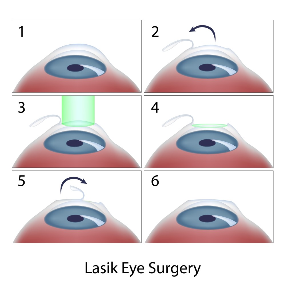
Today, laser in situ keratomileusis (LASIK) is the most popular method of laser eye surgery. LASIK uses an excimer laser to correct nearsightedness, farsightedness, or astigmatism by removing a thin lenslet of tissue from the surface of the cornea (the clear, front “watch crystal” of the eye). This is analogous to removal of a “tissue contact lens”. In LASIK, which is now an “all-laser” technique, a pancake-like thin flap of the cornea is first prepared with a high speed femtosecond laser. The flap acts to preserve the surface epithelial cells (which are like tiles on a floor) to promote quick healing and recovery of vision. Next, the excimer laser is used to remove a small amount of tissue from the corneal surface beneath the flap. The excimer laser used in LASIK produces a beam of invisible ultraviolet light energy, which when applied via an eye tracking mechanism, results in meticulous removal of this “tissue contact lens”. After corneal reshaping, the LASIK procedure is finished when the corneal flap is repositioned. When the flap is replaced, it lies in the bed of excimer laser removed tissue, causing the surface to change shape with the effect of decreasing nearsightedness, farsightedness, or astigmatism.

Laser vision correction can also be performed without a LASIK flap. These procedures, which are also perfomed with the excimer laser, go by a number of names – PRK (photorefractive keratectomy), LASEK (laser epithelial keratomileusis), epi-LASIK, or ASA (Advanced Surface Ablation). Although LASEK and LASIK sound the same, unlike traditional LASIK, LASEK does not require the preparation of a corneal flap. This has two potential advantages. First, risks of making the corneal flap in LASIK are avoided. This may be important in some patients in whom there is an additional risk in making the flap, such as patients with corneal scars or irregularities. Second, since laser treatment is done on the surface, LASEK/PRK preserves more corneal tissue. In particular, patients who have thinner corneas may be more safely treated with a no flap technique rather than LASIK.

At the beginning of the LASEK / PRK eye surgery procedure, the surface cells of the cornea are loosened and removed. The laser treatment then is applied, just as in LASIK, removing the properly shaped “tissue contact lens” for the desired optical correction. At the end of the procedure, a contact lens bandage is applied. Topical drops are used for a few week afterwards to avoid infection and control wound healing.

Vision after LASEK/PRK takes a little while longer to completely improve and stabilize than after LASIK because the epithelium needs to grow and smooth. Substantial improvement usually is noticed the day after the procedure and fluctuates over the next 2 weeks. The contact lens is removed in 5 days in most patients. Driving vision in the days after the procedure can be variable and take up to 2 weeks in some patients.

For both LASIK and LASEK/PRK, there are two basic types of possible side effects. Because patients may respond and heal differently, it is possible that the entire refractive error may not be fully corrected. In this case, vision will be clearer without glasses, but may not be as good as desired. In these situations, patient often can undergo a re-treatment procedure to further improve their vision. In addition, optical side effects include halos around lights and glare, especially at night, and some patients may experience dry eye sensations. Other, more rare, complications include infection or scarring.
LASIK, LASEK, PRK, PTK
Recently, there has been much talk in the keratoconus community about combining corneal collagen crosslinking with topography-guided LASEK/PRK. Topography-guided PRK uses information gained from your corneal map to program the laser to help make your cornea more optically regular. The goal of topography-guided PRK, like Intacs, is to improve corneal contour in the KC patient to improve glasses corrected vision and contact lens tolerance. In general, you will still need contacts and glasses afterwards.

Typically, LASEK/PRK procedures for keratoconus are combined with corneal collagen crosslinking, which has the goal to strengthen the weak keratoconic cornea and decrease progression of corneal mishapening over time. It is important to note that such treatments are not FDA-approved and are not generally available in the U.S. However, a number of international surgeons have been exploring the potential role of combined LASEK/PRK with crosslinking to improve keratoconus outcomes. In our practice, we have also had the opportunity to use Intacs and other procedures to further improve corneal shape in patients who have undergone topography-guided treatments with crosslinking with encouraging results.

In addition to LASIK and LASEK/PRK, the excimer laser may provide a novel therapeutic modality in the treatment of a number of superficial corneal disorders. This treatment is known a phototherapeutic keratectomy or PTK. Whether PTK eye surgery is used alone or as an adjunctive strategy in traditional corneal surgical techniques, a number of disorders affecting the corneal surface may be successfully treated by taking advantage of the excimer laser’s ability to meticulously remove superficial corneal tissue. These include a variety of corneal degenerations and dystrophies, corneal irregularities, and superficial scars, such as surface nodules found at the apex of the keratoconic cone. While some of these conditions, heretofore, could be treated by mechanical superficial keratectomy techniques, PTK may minimize tissue removal and surgical trauma.

So, for patients with keratoconus, it is important to know that, although LASIK type procedures are generally not indicated, research using these advanced technologies continues. A tailored therapeutic approach over time may combine a variety of procedures to optimize the corneal shape and ultimate visual outcome for the patient with keratoconus.

9/15/15


Peter Hersh, MD - Laser Refractive Surgery: LASIK, LASEK, PRK and PTKPeter S. Hersh, MD
Cornea and Laser Eye Institute – Hersh Vision Group
CLEI Center for Keratoconus

Which Eye Care Specialist Do You Need?

It’s time to get your eyes checked – do you go to an ophthalmologist, optometrist or optician? Your optometrist sees the beginnings of age-related macular degeneration, but is sending you to see and ophthalmologist, why?

One of the most confusing things about taking care of your eyes can be differentiating between an ophthalmologist, optometrist and optician. Each eye care specialist has a very important part to play in the health of your eyes and here is a quick synopsis of what each does so you can choose the best one for your vision issues and treatment.

Ophthalmologist
These specialists are fully trained medical doctors that have completed the eight years of training beyond a bachelor’s degree. Their training has included a full spectrum of eye care, from prescribing glasses and contact lenses and giving eye injections, to carrying out intricate eye surgeries. Many doctors may also be involved eye research to better understand vision, improve eye disease treatments or potentially find a cure. They are easily identified by the MD following their name.

Optometrist
These medical professionals have completed a four-year program at an accredited school of optometry. They have been trained to prescribe and fit glasses and contact lenses, as well as diagnose and treat various eye diseases. They provide treatments through topical therapeutic agents and oral drugs, and are licensed to perform certain types of laser surgery, such as Lasik. They are easily identified by the OD following their name.

Optician
These eye care professionals are not licensed to perform eye exams, medical tests or treat patients. Their purpose is to take the prescription from the ophthalmologist or optometrist and work with you to determine which glasses or contact lenses work best for you. If you suffer from an eye disease like keratoconus, these specialists can make the difference between a relatively normal life, or one that is dictated short periods of vision because of contact lens pain. These eye care professionals may hold and associate optician degree or have apprenticed fore required number of hours.

While each one of these eye specialists has their own area of expertise, they can form a team whose only concerns are your eye health and the ability to see as clearly as possible.

8/11/15


Susan DeRemerSusan DeRemer, CFRE
Vice President of Development
Discovery Eye Foundation

Eye Issues For Every Age Recap

Vision is something we take for granted, but when we start to have trouble seeing it is easy to panic. This blog has covered a variety of eye issues for every age, from children through older adults. Here are a few articles from leading doctors and specialists that you may have missed and might be of interest.
Artistic eye 6
Bill Takeshita, OD, FAAO – Visual Aids and Techniques When Traveling

Michelle Moore, CHHC – The Best Nutrition for Older Adults

Arthur B. Epstein, OD, FAAO – Understanding and Treating Corneal Scratches and Abrasions

The National Eye Health Education Program (NEHEP) – Low Vision Awareness
Maintaining Healthy Vision

Sandra Young, OD – GMO and the Nutritional Content of Food

S. Barry Eiden, OD, FAAO – Selecting Your Best Vision Correction Options

Suber S. Huang, MD, MBA – It’s All About ME – What to Know About Macular Edema

Jun Lin, MD, PhD and James Tsai, MD, MBA – The Optic Nerve And Its Visual Link To The Brain

Ronald N. Gaster, MD FACS – Do You Have a Pterygium?

Anthony B. Nesburn, MD, FACS – Three Generations of Saving Vision

Chantal Boisvert, OD, MD – Vision and Special Needs Children

Judith Delgado – Driving and Age-Related Macular Degeneration

David L. Kading OD, FAAO and Charissa Young – Itchy Eyes? It Must Be Allergy Season

Lauren Hauptman – Traveling With Low Or No Vision  /  Must Love Dogs, Traveling with Guide Dogs  /  Coping With Retinitis Pigmentosa

Kate Steit – Living Well With Low Vision Online Courses

Bezalel Schendowich, OD – What Are Scleral Contact Lenses?

In addition here are few other topics you might find of interest, including some infographics and delicious recipes.

Pupils Respond to More Than Light

Watery, Red, Itchy Eyes

10 Tips for Healthy Eyes (infographic)

The Need For Medical Research Funding

Protective Eyewear for Home, Garden & Sports

7 Spring Fruits and Vegetables (with some great recipes)

6 Ways Women Can Stop Vision Loss

6 Signs of Eye Disease (infographic)

Do I Need Vision Insurance?

How to Help a Blind or Visually Impaired Person with Mobility

Your Comprehensive Eye Exam (infographic)

Famous People with Vision Loss – Part I

Famous People with Vision Loss – Part II

Development of Eyeglasses Timeline (infographic)

What eye topics do you want to learn about? Please let us know in the comments section below.

7/21/15


Susan DeRemerSusan DeRemer, CFRE
Vice President of Development
Discovery Eye Foundation