Chlorine & Your Eyes

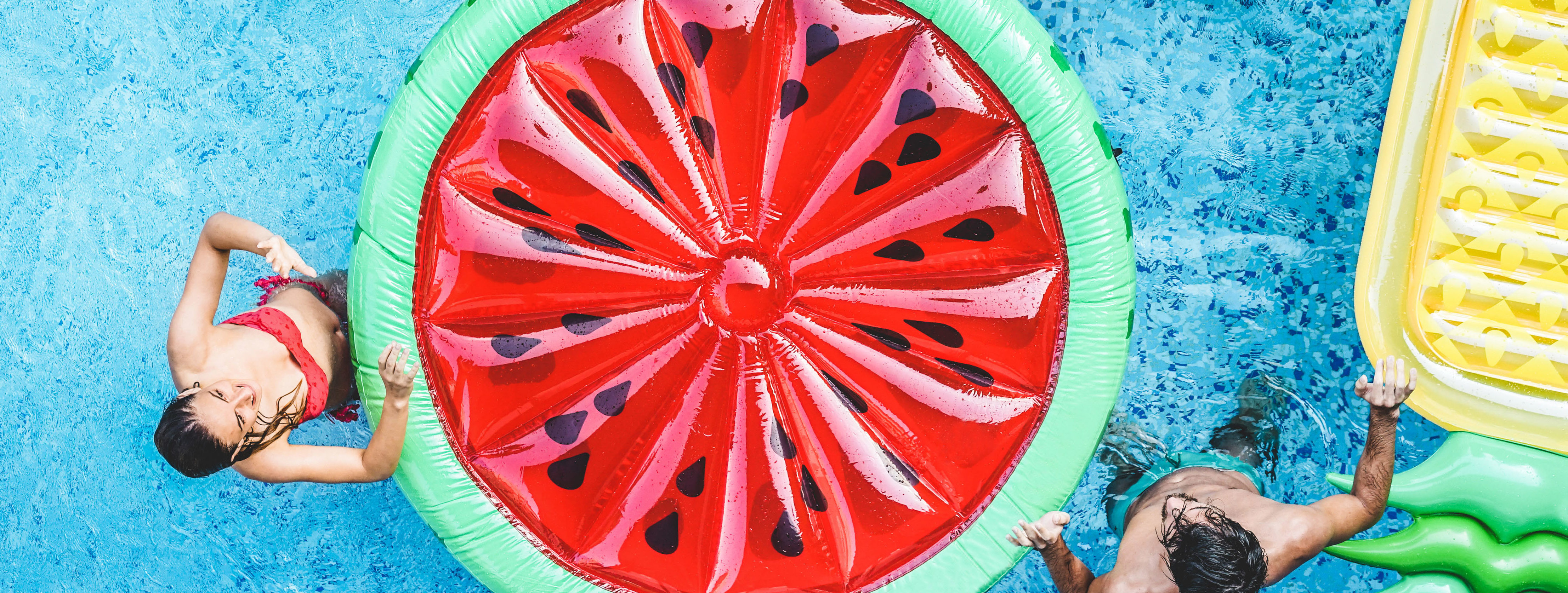
Summer time is officially here and everyone enjoys a dip in a nice, cool pool during the summer months.  While swimming is a great form of exercise and a relaxing way to cool down, the water can be hard on your eyes.

What Chlorine Does to Your Eyes –

Yes chlorine can make your eyes red!  But the real dangers in pool water aren’t just a result of your eyes coming in contact with the chemical. The redness and discomfort that sometimes accompany swimming can be caused by bacteria that linger in the water. When submerged in chlorine-treated water, your eyes lose the tear film that protects against infection.  Even though the purpose of chlorine in pools is to reduce the amount of harmful bugs, some contaminants are resistant to the chlorine that is used. This means the health of your eyes can be compromised with infections caused by bugs still lingering in the water.  

This can result in these three common eye issues:

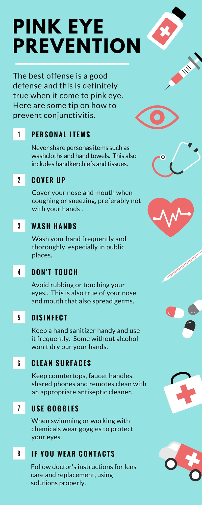
  • Pink eye or conjunctivitis – This is one of the most common eye infections swimmers can get, as it can be either viral or bacterial and spreads quickly and easily through the water.
  • Red, irritated eyes –  This is a result of your eyes becoming dehydrated due to the chlorine and the removal of your tear film. Sometimes you may also experience blurriness and distorted vision, this is usually only temporary.
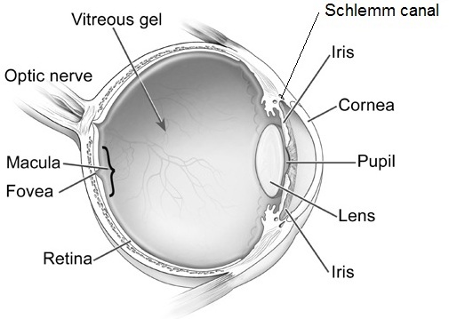
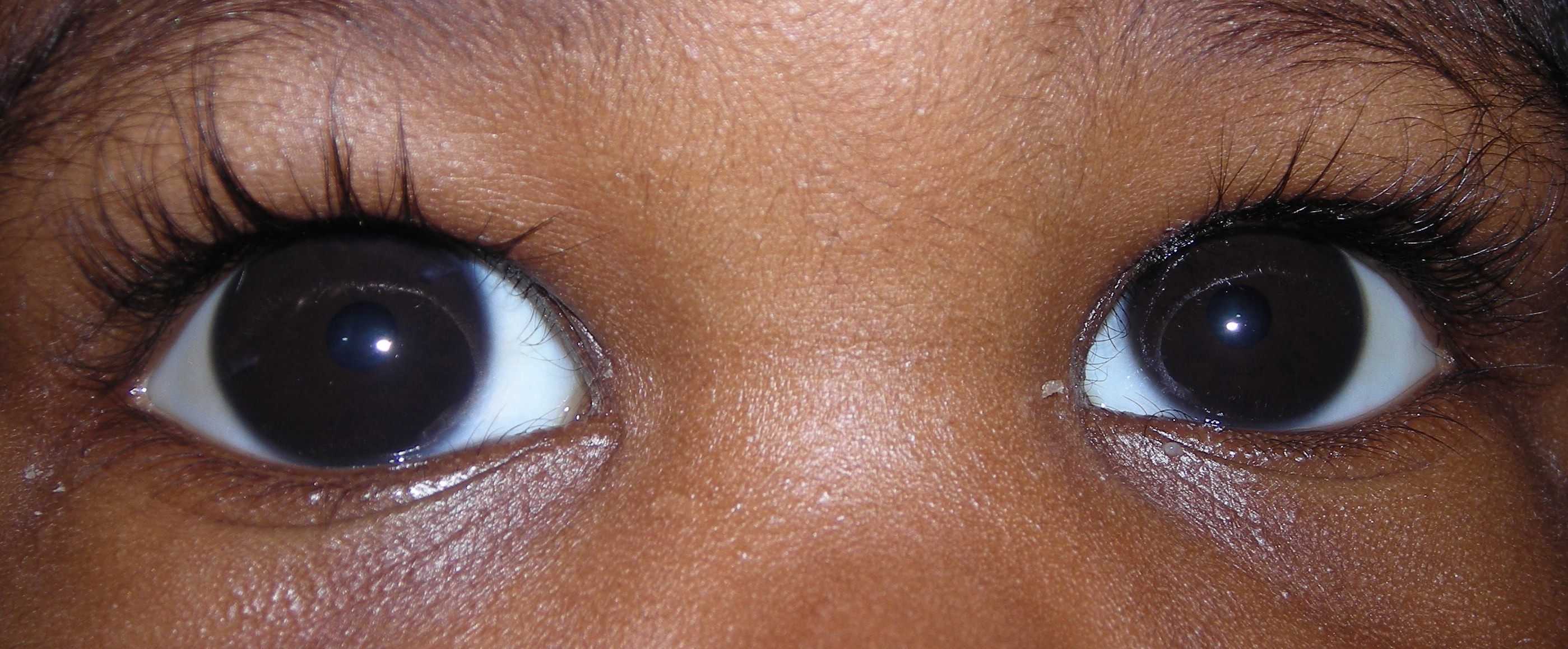
  • Acanthamoebic keratitis – This is a severe eye infection that is caused by amoeba in the water becoming trapped between the cornea and the contact lens. It can begin to live there, which can result in ulcers on your cornea and permanent damage to your vision.


Swimming with Contact Lenses  

Wearing contact lenses in any type of water—including a pool, hot tub, ocean or lake—puts you at higher risk for a corneal infection. Bacteria and other microbes can grow on the lenses even after just one swim. Because contact lenses sit on your eyes for an extended period of time, your eyes are then continuously exposed to chemicals, bacteria, fungi or parasites after you swim. That can lead to a painful infection, corneal damage, and even loss of vision.

To avoid any kind of infection, remove contacts altogether when swimming or use swim goggles. You can get prescription swimming goggles to help keep your vision clear and eyes healthy in the pool. Talk with your eye care provider for more information about the different kinds of swim goggles available.

If you have any of these eye infection symptoms are increasing one hour or longer after swimming, see your eye doctor right away.

Redness
Pain
Tearing
Being very sensitive to light
Blurry vision
Sensation of having something in your eye
Discharge from your eye
Eye swelling
 

With all of these risks to your eyes from chlorine, swimming might seem a bit scarier than before. However, there is no need to panic!  By taking a few safety measures, you can protect your eyes and still enjoy your time in the pool.


  • Wear Goggles – Wear a pair of swim goggles every time you swim. Goggles keep pool chemicals out of your eyes.


  • Wash Your Eyes – Immediately after swimming, splash your closed eyes with fresh tap water.  This washes chlorine and other chemicals off your eyelids and eyelashes.


  • Use Eye Drops – Use over-the-counter lubricating eye drops before and after swimming to keep the tear film balanced and eyes comfortable.


  • or Use Gel Tears – If you have dry eye, thicker artificial tears called gel tears will help protect your tear film, use these drops before putting on your goggles.


  • Stay Hydrated – Don’t forget to drink plenty of water. Staying well hydrated is an important part of keeping your eyes moist and comfortable.


Don’t miss out on the fun this summer! By taking these easy steps whenever you decide to take a dive, you can have peace of mind that your eyes and your vision are protected.


April is Sports Eye Safety Month

Basketball, Baseball and Air/Paintball Guns Top the List of Leading Causes of Eye Injuries

More than 40 percent of eye injuries that occur every year are related to sports or recreational activities. A recent study found that about 30,000 people in the U.S. went to an emergency department with a sports-related eye injury, a substantially higher estimate than previously reported. Three sports accounted for almost half of all injuries: basketball, baseball and air/paintball guns. 

Basketball was the leading cause of injury in males, followed by baseball/softball, and air/paintball guns. Baseball or softball was the leading cause among females, followed by cycling and soccer.

In support of Sports Eye Safety Month in April, we are offering athletes of all ages guidance on how to protect their eyes.

Sports-related injuries can range from corneal abrasions and bruises on the lids to more serious, vision-threatening internal injuries, such as a retinal detachment and internal bleeding. About one-third of sports related eye injuries happen to kids.

The good news is that simply wearing protective eyewear can prevent about 90 percent of eye injuries. Follow these tips to save your vision:

  • Wear the right eye protection: For basketball, racquet sports, soccer and field hockey, wear protection with shatterproof polycarbonate lenses.
  • Put your helmet on: For baseball, ice hockey and lacrosse, wear a helmet with a polycarbonate face mask or wire shield.
  • Know the standards: Choose eye protection that meets American Society of Testing and Materials (ASTM) standards. See the Academy’s protective eyewear webpage for more details.
  • Throw out old gear: Eye protection should be replaced when damaged or yellowed with age. Wear and tear may cause them to become weak and lose effectiveness.
  • Glasses won’t cut it: Regular prescription glasses may shatter when hit by flying objects. If you wear glasses, try sports goggles on top to protect your eyes and your frames.

Virtually all sports eye injuries could be prevented by wearing proper eye protection. That is why athletes are encouraged to protect their eyes when participating in competitive sports.

Anyone who experiences a sports eye injury should immediately visit an ophthalmologist, a physician specializing in medical and surgical eye care.


Back to School – Why Eye Exams are Important!

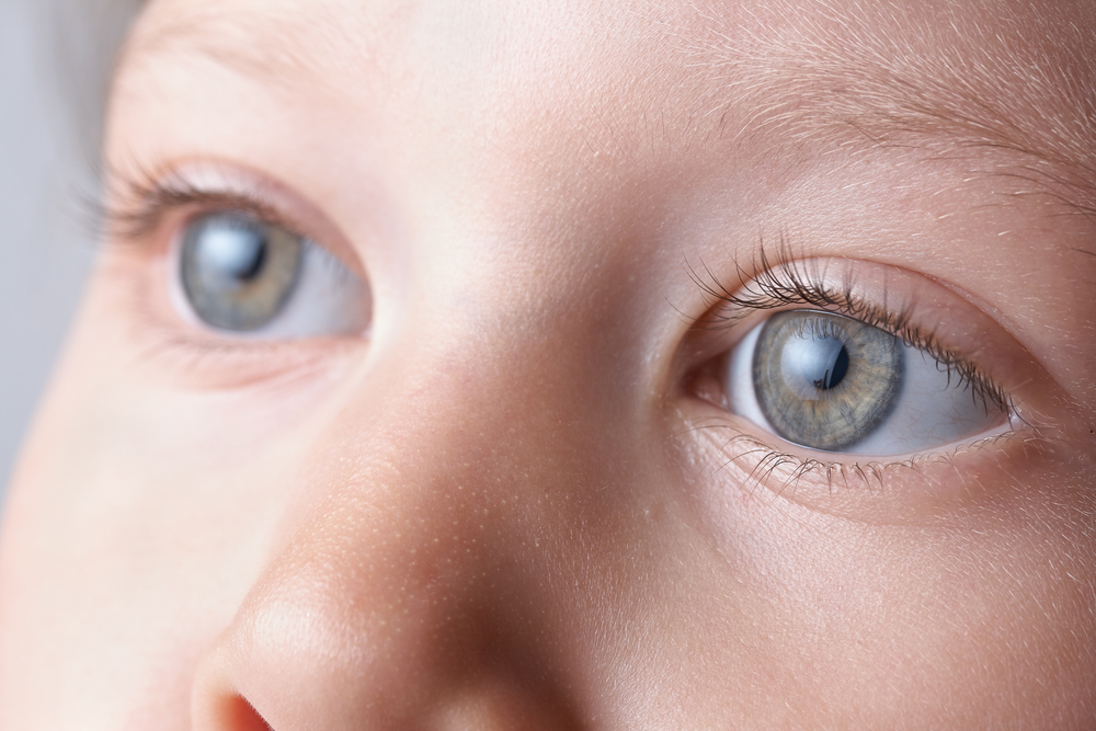
Summer is almost over and it’s back to school season. As parents, many of us are busy ensuring our kids are ready and prepared for the new year; worrying about school supplies, new clothes, and new haircuts. There is always a long list of things to do before school starts. But something that often gets overlooked is getting your child’s eyes examined annually.

Early eye examinations are crucial to make sure children have normal, healthy vision so they can perform better at schoolwork and play. Early identification of a child’s vision problem can be crucial because children often are more responsive to treatment when problems are diagnosed early.

Early eye exams also are important because children need the following basic skills related to good eyesight for learning:

  • Near vision

  • Distance vision

  • Binocular (two eyes) coordination

  • Eye movement skills

  • Focusing skills

  • Peripheral awareness

  • Hand-eye coordination

Parents also need to be alert for the presence of vision problems such as ‘crossed’ eyes or ‘lazy’ eye. These conditions can develop at a young age. ‘Crossed’ eyes or strabismus involves one or both eyes turning inward (towards the nose) or outward. Amblyopia, known as ‘lazy’ eye, is a lack of clear vision in one eye, which can’t be fully corrected with eyeglasses. ‘Lazy’ eye often develops as a result of ‘crossed’ eyes, but may occur without noticeable signs. Lazy eye can be treated if caught early.

In addition, parents should watch their child for indication of any delays in development, which may signal the presence of a vision problem. Difficulty with recognition of colors, shapes, letters and numbers can occur if there is a vision problem. Children generally will not voice complaints about their eyes, therefore parents should watch for signs that may indicate a vision problem, including:

  • Sitting close to the TV or holding a book too close

  • Squinting

  • Tilting their head

  • Constant eye rubbing

  • Extreme light sensitivity

  • Poor focusing

  • Poor visual tracking (following an object)

  • Abnormal alignment or movement of the eyes (after 6 months of age)

  • Chronic redness of the eyes

  • Chronic tearing of the eyes

  • A white pupil instead of black

Scheduling Eye Exams for Your Child

If eye problems are suspected during routine physical examinations, a referral should be made to an eye doctor for further evaluation. Eye doctors have specific equipment and training to assist them with spotting potential vision problems in children.

When scheduling an eye exam for your child, choose a time when he or she usually is alert and happy.

Glasses and Contacts

Keep these tips in mind for kids who wear glasses:

  • Plastic frames are best for children younger than 2.

  • Let kids pick their own frames.

  • If older kids wear metal frames, make sure they have spring hinges, which are more durable.

  • An elastic strap attached to the glasses will help keep them in place for active toddlers.

  • Kids with severe eye problems may need special lenses called high-index lenses, which are thinner and lighter than plastic lenses.

  • Polycarbonate lenses are best for all kids, especially those who play sports. Polycarbonate is a tough, shatterproof, clear thermoplastic used to make thin, light lenses. However, although they’re very impact-resistant, these lenses scratch more easily than plastic lenses.

  • Your eye doctor can help you decide what type of vision correction is best for your child.

Specialists state that 80% of what your youngster learns in school is taught visually. Untreated vision troubles can put children at a substantial disadvantage. Be certain to arrange that your child has a complete eye exam before school starts.

View Video

Issues That Could Affect Your Child’s Vision

boy-with-glassesVision loss is feared more than the loss of any other sense and is considered to affect the quality of life more than most other issues. When it comes to children, even partial vision loss can be damaging because it can affect the way that your child learns and develops. There are several different types of issues that may affect your child’s vision. Awareness is key to prevention and treatment.

Refractive Errors

Refractive errors such as nearsightedness, farsightedness, and astigmatism are the most common types of issues that affect children’s vision. Since children are working to constantly adapt to their surroundings, they may not realize that they have a vision problem and the issue may manifest as an inability to focus, chronic headaches, or poor eye-hand coordination. In most cases, these issues can be corrected with glasses or contacts, but extreme cases may require surgery.

Alignment Disorders

Alignment disorders are generally more obvious than other types of issues. One eye may drift to the side, an eyelid may droop, or the surface of an eye may appear cloudy, affecting the vision. In some cases, however, the condition may be intermittent, so it is important to continually look for these symptoms and alert an eye doctor to any concerns. Alignment disorders may be corrected with surgery, an eye patch, eye drops, or a combination depending on the cause.

Pediatric Retinoblastoma

Pediatric retinoblastoma is a type of kid’s eye cancer that usually affects children under six years of age. About 95 percent of children diagnosed in the US are able to be treated successfully and a majority of these children retain most or all of their sight. The prognosis for retinoblastoma improves greatly with early diagnosis and treatment. One of the most common warning signs that you can look for is a white glare on the pupil when it is directly exposed to light.

Diseases and Infections

Diseases and infections such as conjunctivitis, styes, and blocked tear ducts are usually minor problems that are easily resolved. However, these issues may develop into larger problems that affect the vision if care is not taken. Conjunctivitis may resolve on its own depending on the cause, but the child should be kept away from others during the healing process to avoid infecting others or being exposed to other contaminants while the eye is sensitive.

Blocked tear ducts may be opened up using massage techniques recommended by a pediatrician or ophthalalmogist. Allowing the eyes to dry out may be dangerous for the vision, so drops may be needed to keep the eye moist while the tear ducts are blocked. Styes are caused by an infection in the eyelash follicle, so it is important to keep the area clean so that the infection can clear without causing damage.

By working to spot potential issues, you can help to preserve your child’s eye health and vision.


amanda-duffyAmanda Duffy
Freelance blogger

Pink Eye Tips and Prevention

pink eye
Pink eye is an inflammation or infection of the thin, clear covering of the white of your eyeball (the conjunctiva) and the inside of your eyelids. When the small blood vessels in the conjunctiva become inflamed they are more visible making the whites of your eye to appear pink. Also called conjunctivitis, it can affect one or both eyes.

Common symptoms of pink eye include:

  • Redness in the white of the eyeball(s) and or inner eye lid(s)
  • Increased tearing or discharge
  • Slightly blurred vision from discharge
  • Crusting of the eyelashes from the discharge that may prevent eyes from opening after sleep
  • Mild eyelid swelling
  • Itching or burning sensation
  • Increased sensitivity to light
  • Irritation or gritty feeling in your eye(s)

Make an appointment with your eye doctor if you notice and of the symptoms of pink eye. Some forms are highly contagious for as long as two weeks, so an early diagnosis could protect those around you from contacting the disease. If you were contact lenses, stop using them until directed by your doctor.

There are four general types of pink eye.

Allergic Conjunctivitis
This form is caused by eye irritants such as pollen, dust, animal dander and other environmental factors. It is not contagious. Treatment often includes applying a cool compress to your eyes and using allergy eye drops and artificial tears. In severe cases non-steroidal and anti-inflammatory medications may be prescribed.

Bacterial Conjunctivitis
This type is most often caused by staphylococcal or streptococcal bacteria, is highly contagious and can cause serious damage to the eye if left untreated. This is treated with antibiotic eye drops or ointments to speed up the healing process that can take one to two weeks. While you may see improvement after three to four days, the entire course of treatment needs to be used to prevent a recurrence.

Because this is so highly contagious here are a few things to remember so you don’t spread it to others or re-infect yourself:

  • Don’t touch your eye with your hands
  • Wash your hands frequently and thoroughly
  • Change towels and washcloths daily – and don’t share them
  • Change pillowcases often
  • Get rid of all eye cosmetics and personal care items such as eye creams – and don’t share them
  • Avoid swimming
  • Don’t reuse tissues when wiping your eyes, and throw them out immediately
  • Follow your eye doctor’s instructions related to your contact lens usage and care

Viral Conjunctivitis
This is the same type of virus associated with the common cold. Antibiotics will not work on a viral infection. Like a cold, the infection just needs to run its course which could take anywhere from a few days up to 2-3 weeks. It is also contagious like a cold, so follow the same instructions as listed above to not spread the infection.

Chemical Conjunctivitis
This can be caused by irritants like air pollutions, chorine in swimming pools or exposure to noxious chemicals. To treat this type of pink eye requires a doctor to carefully flush your eyes with saline and may require topical steroids. Acute chemical injuries are very serious and need prompt medical attention to avoid corneal scarring, intraocular damage, vision loss or the loss of an eye.

Of course the best way to deal with pink eye is not to get it. Here are some ways to protect yourself and others.
pink eye

2/24/16

Susan DeRemerSusan DeRemer, CFRE
Vice President of Development
Discovery Eye Foundation

Accommodative Esotropia

This article is reprinted with the permission of Dr. Kenneth W. Wright, Medical Director of the Wright Foundation for Pediatric Ophthalmology & Strabismus. Check out his website for information on over 20 pediatric eye disease and conditions.
Accommodative Esotropia

Normal Binocular Vision

Normally, both eyes are aligned on the same visual target and the images from each eye are merged in the brain to form a single three-dimensional image, or binocular vision. The brain’s process of merging or “fusing” images from each eye into one image is called binocular fusion. The perception of three-dimensional depth is called stereoscopic vision. Stereoscopic vision is the highest level of binocular vision and requires intricate processing of information from both eyes. Binocular vision develops during early infancy, and proper alignment during this time is necessary for normal binocular development to occur.

Accommodative Esotropia

Esotropia means one eye is turned in towards the nose, or crossed eyes. Patients with esotropia have one eye aligned on the visual target, but the other eye is turned in towards the nose. When the eyes are crossed only one eye is aligned with the target and the child is forced to use only one eye for vision. Accommodative esotropia is a type of esotropia caused by significant farsightedness (hypermetropia). Most think that farsighted people can see well only in the distance. In children, this is not true. Children have the ability to focus great amounts, so most children can see well for distance and near even without glasses. Focusing (medically termed accommodation) is the process of increasing the lens power of the eye to see clearly. Linked to focusing is the natural reflex of convergence (eyes move in). As one focuses on an approaching near object, the eyes move in to stay on target. This process of focusing and convergence used for near vision is termed the near reflex. Accommodative esotropia occurs because the farsighted child has to over focus to see clearly. When eyes over focus, the natural reflex is for the eyes to cross. You can experience this by trying to see the tip of your nose. When you look at the tip of your nose you have to over focus and consequently your eyes cross. Since more focusing is needed to see near objects, the crossing tends to be greater when looking at close objects.

Accommodative Esotropia eye turns
Right eye turns in because patient is farsighted and not wearing glasses.
Accommodative Esotropia corrected
Eyes are in excellent alignment after prescribing glasses.


The onset of accommodative esotropia is most commonly seen between the ages of 2 and 4; however, even infants can have crossed eyes. This situation is usually first noticed when the child is tired, sick, or looking at an object very near to their face. Some children cross when they are tired and this is because they cannot sustain the effort to keep the eyes straight. The crossing is usually intermittent at first, but can quickly become constant. There may be a parent or a close relative with the same problem; however, in many cases there is no family history of crossed eyes.

During the examination, three important determinations are necessary. The first determination is to make sure the vision is normal in each eye. This is done by assessing the visual behavior of the preverbal child, or simply having the verbal child read the eye chart. Secondly, the amount of crossing is evaluated. This is measured using prisms while the child is viewing an object. Thirdly, the need for glasses is measured, and this requires drops to dilate the pupil and relax the child’s focusing. These drops take approximately 20 to 30 minutes to work and will blur vision for 1 to 3 hours, but the pupil may stay large for much longer. After the eyes have been dilated, the eyeglass prescription is calculated using a special light (retinoscope) along with lenses. Determining the proper lens power in young children is difficult and may require repeat exams and changes in the eyeglass lenses.

Effects of Esotropia on Visual Development

Esotropia occurring in young children and infants results in the immature brain turning off the information from the deviated eye. This mechanism of turning off visual areas of the brain connected to the deviated eye is called “suppression.” Thus, patients with esotropia use one eye at a time (monocular vision) and do not have binocular fusion or stereoscopic vision. Suppression disrupts normal binocular visual development and if not treated early, causes permanent loss of binocular vision and stereoscopic vision. Early treatment of esotropia is critical to stimulate binocular development.

How Do Patients with Esotropia See?

If the esotropia is acquired in late childhood (after 7 to 9 years of age) or in adulthood, it will cause double vision. Esotropia occurring in infants and young children, however, does not cause double vision, as the young, immature brain has the ability to suppress the information from the deviated eye. The child uses one eye at a time to see and avoids double vision. The fact that the eyes are crossed disrupts normal binocular visual development and often causes permanent loss of binocular vision and stereoscopic vision. Early treatment of esotropia is critical in order to stimulate binocular development.

Treatment of Accommodative Esotropia

Glasses

The goal of treatment is to align the eyes, stimulating them to work together to establish binocular vision and stereoscopic vision. Children and infants who are significantly farsighted are best treated with glasses. If the glasses align the eyes, then surgery is not necessary, and the treatment is to continue with the glasses. The full, hypermetropic (farsighted) prescription is usually given via eyeglasses, and most parents are surprised at how well these children adjust to the glasses. When properly worn, most children adapt to the glasses like “fish to water.” The glasses not only straighten the eyes, but also relax the child’s vision, as they no longer have to over focus. In patients with accommodative esotropia, glasses must be worn full-time. Older children over 4 to 5 years may have blurred distance vision when they first put on their glasses. This is because they had a strong habit of over-focusing and continue to do so even when wearing the glasses. Over several days, most children will relax their over-focusing and enjoy the comfort the glasses afford. In those children who do not adapt to the glasses, drops can be used to relax focusing, or a reduced prescription power can be given. In most cases, however, the best treatment is to give the full power. The eyes usually straighten within a few days to a few weeks after wearing the glasses. If the eyes are still crossing with the glasses and the child is not using the eyes together after several weeks, then eye muscle surgery is usually required. Occasionally, an initial response to glasses is that the eyes “break down” and cross for distance and near. In this situation, surgery in addition to the glasses may be required.

Bifocal Glasses

In certain children, glasses will align the eyes for distance viewing, but the eyes will still cross for near work. These patients can be helped with bifocal glasses. A bifocal is a small powerful lens placed in the lower part of the eyeglass lens. This more powerful lens will further relax near focusing to straighten the eyes for near work. Chin-up posturing for near work indicates that the child is using the bifocals correctly and is viewing through the bifocal lens for near work.

Amblyopia

Some children with crossed eyes have a strong fixation preference for one eye (dominant eye) and constantly have one eye turned in. Constant use of only one eye can lead to vision loss of the deviated, or non-dominant, eye. Poor vision occurs as visual areas in the brain connect with the dominant eye and are then subsequently suppressed. If left untreated, the deviated eye will progressively lose vision over time. This poor vision caused by brain suppression is called amblyopia. Amblyopia occurs only in young children when the visual areas are immature and still developing. Children who have a difference in the strength of the glasses in one eye as compared to the other have an increased risk of developing amblyopia in the more farsighted eye. Approximately 20 to 40% of patients with esotropia will also have amblyopia of the non-preferred eye.

Patching

If amblyopia is present, patching of the good eye is indicated to promote visual stimulation of the amblyopic eye and improve vision. Patching does not straighten the eyes and is not indicated if vision is equal. Another way to promote stimulation of the amblyopic eye is to blur the vision of the “good eye.” This can be done by placing a blurring lens over the good eye, or by administering drops to blur the good eye. In most cases, patching the good eye with an adhesive patch is the most practical treatment. Patching is continued until vision improves in the weaker eye, usually taking a few weeks to several months. In the vast majority of patients, vision can be improved if the parents and child are compliant with the treatment.

10/8/15

Dr. Kenneth WrightKenneth W. Wright, MD
Medical Director, Wright Foundation for Pediatric Ophthalmology & Strabismus
Clinical Professor of Ophthalmology, USC Keck School of Medicine

Primary Congenital Glaucoma

What is primary congenital glaucoma?

Glaucoma in children includes a variety of disorders in which drainage system of the eye does not function adequately, leading to abnormally high pressure inside of the eye (the intraocular pressure, or IOP), and resulting in damage to many different structures of the child’s eye. If not treated promptly and successfully, pediatric glaucoma can lead to severe vision loss or even blindness in one or both eyes. In primary childhood glaucoma, the drainage system usually has not formed properly (often resulting from a genetic abnormality) while in secondary childhood glaucoma, the abnormal fluid outflow problem results from other problems with the eye(s), sometimes accompanied by other medical problems outside the eyes.

Primary congenital glaucoma is the most common of the primary childhood glaucoma types, although it is still rather rare. Let’s take a moment now to review the parts of the eye, and eye’s drainage system, sometimes also called the “aqueous outflow pathway”, since it drains the fluid within the eye (the aqueous humor), which is separate from the tears that flow on the outside of the eye’s surface and then into the nose or down a child’s cheeks.

The aqueous outflow pathway of the eye (comprising both the trabecular meshwork and Schlemm canal), situated at the junction (or “angle”) between the iris (the colored portion of the eye) and the sclera (the white part of the eye), has not formed correctly (Figure 1).

primary congenital glaucoma
Figure1. Schematic eye shows different structures of the ocular globe. Note that the Schlemm canal is part of the drainage system of the eye . Modified from National Eye Institute.

The aqueous humor therefore builds up within the front portion of the eye, causing abnormal elevation of the IOP.

In contrast to the eyes of adults and older children, the entire eye in infants and young children is distensible and the high IOP in primary congenital glaucoma often causes stretching and damage to several parts of the eye; this most often results in enlargement, clouding and scaring of the cornea (the front window of the eye) as well as severe nearsightedness, damage to the optic nerve, and resulting poor vision.

Primary congenital glaucoma (also called PCG) is almost always genetic, although usually there is no one else in the family with the condition. It is not related to anything that the parents did (or did not do) during the pregnancy or afterwards, and does not have any relationship to the baby’s sex or racial background. It occurs in about 1 every 10,000 to 20,000 births in western countries, but may be more common in certain populations of the world. Most babies with this disease are otherwise normal.

How is primary congenital glaucoma diagnosed?

Most cases present within six months of birth, with nearly 80% presenting before one year of age. In 70- 80 % of cases both eyes are affected. Most cases present for medical attention due to the size or cloudy appearance of the cornea in one or both eyes (Figure 2).

primary congenital glaucoma
Fig 2. Left eye of child with congenital glaucoma. The eye is enlarged and the front part of the eye is cloudy (corneal edema).

In cases where only one eye is affected, a difference in size can be seen between the two eyes and this sometimes brings the baby to the ophthalmologist (Figure 3).

primary congenital glaucoma
Fig 3. Different size of eyes in a child with congenital glaucoma. Note the increased size of the right eye. The brown area (iris) and the transparent part in front of the color part (cornea) are significantly larger in the right eye.

The diagnosis of PCG is based on clinical findings and there are three classic signs that the child can present with:

  • abnormal sensitivity or intolerance to light (photophobia)
  • excessive blinking or squinting of the eyelids (blepharospasm)
  • excessive tearing (epiphora)

The exam in clinic can be challenging for infants and young children and most require an exam under anesthesia, to allow detailed examination of the eye(s) that would not be possible in the clinic. Often the ophthalmologist will be able to follow the examination under anesthesia with the most appropriate surgery for the glaucoma, if surgery is indeed required.

How is primary congenital glaucoma treated?

PCG is almost always treated with surgery, although medications are often needed to help in addition to the surgery. Medications are very useful before initial surgery to help reduce the IOP and decrease the clouding of the cornea. In addition, medications may be recommended to keep the IOP to a safe level after surgery has been performed. If the IOP is not controlled successfully, or if damage has been substantial prior to diagnosis and treatment, PCG causes severe vision loss and can even cause blindness. Sometimes the damage from PCG is uneven between a child’s two eyes, leading to amblyopia (“lazy eye”) in the more severely affected size.

The initial surgical procedure of choice is usually aimed at opening the trabecular meshwork and Schlemm canal (the aqueous outflow pathway) of the affected eye(s). This so-called “angle surgery” can be performed either from inside of the eye (goniotomy) or externally (trabeculotomy), and may need to be repeated more than once in some cases.

If angle surgery fails, other procedures are available to allow the aqueous humor fluid to exit the eye (glaucoma drainage device or filtration surgery), or even to decrease the amount of fluid the eye makes (cycloablation procedures). For these more difficult procedures, the child is usually referred to an ophthalmic surgeon with expertise in treating childhood glaucoma.

What is the prognosis for children with primary congenital glaucoma?

While vision loss can be severe, prompt diagnosis and effective treatment and follow-up for children with PCG usually allows affected children to have best-corrected vision of at least 20/50 vision in their better-seeing eye. Children with PCG require continued careful follow-up and treatment their lifetime, and may require more than one surgery, eye drops, and spectacles.

Successful care for children with PCG takes a dedicated team including the family, ophthalmologist, teacher and community support, and the child him/herself.

10/1/15

primary congenital glaucomaElena Bitrian, MD
Assistant Professor of Ophthalmology, Division of Glaucoma
Mayo Clinic


primary congenital glaucomaSharon F Freedman, MD
Professor of Ophthalmology and Pediatrics
Chief of Pediatric Ophthalmology
Duke Eye Center, Duke University

Cortical Visual Impairment: What Is It?

What Is It?

Cortical Visual Impairment (CVI) refers to decreased vision resulting from the visual processing parts of the brain (e.g., the posterior visual pathways and/or the occipital lobes) rather than from the eyes themselves. For example, individuals with CVI typically have normal eye exam findings. However, vision loss from CVI can range from mild to total blindness. It is also one of the more frequent causes of visual impairment in children. Regardless, children with CVI often have some level of vision that may improve over time, particularly if they receive therapy to teach them how to integrate the visual signals their brains are receiving.
cortical visual impairment
CVI may be caused by a number of different conditions that damage the visual parts of the brain. Examples include stroke, decreased blood supply to the brain, decreased oxygenation in the brain, brain malformation or infection, hydrocephalus, seizure, metabolic diseases, head trauma, and other neurologic disorders. Conditions such as these make it difficult for the brain to understand and interpret what the eyes see.

In most cases, individuals with CVI do not have other neurological problems, although epilepsy and cerebral palsy are not uncommon. The presence of CVI is not an indicator of the cognitive abilities of the individual; therefore, CVI should be distinguished from vision loss secondary to global neurological damage, where other functional deficits are also present in motor, cognitive, and physical abilities.

Indicators of Cortical Vision Loss

In children, one of the most common indicators of CVI is their poor attention to visual targets, particularly to more complex targets, such as a person’s face. Other indicators are that children with CVI often prefer to look at lights for long periods of time and that when reaching for an object, they will often look away from the object before grabbing it. This is because children with CVI have difficulty integrating visual stimuli (looking at an object) with their motor ability (grabbing the object). The diagnosis of CVI is given based on the combined results of magnetic resonance imaging (also known as an MRI) and an eye evaluation by a pediatric ophthalmologist.

Treatments

If a child is suspected of having CVI, he or she should be evaluated by a pediatric ophthalmologist as part of the initial evaluation. The pediatric ophthalmologist will assess the child’s eye health as well as the need for glasses to make sure there are no additional factors that may be limiting vision.

Often, there is concern that a child with CVI has little to no vision early in life. However, vision is a learned sense, so as the child matures, he or she may have improved visual responses. As such, early intervention is important for improved visual responses over time, as well as because the treatment period for visual development is limited to the early years of life. State and local educational agencies and early intervention programs should be contacted as soon as a visual concern is noted so that an organized plan of visual stimulation activities can be developed and implemented, based on the specific needs of each child. The professionals involved in the evaluation of a child with vision loss from CVI can include teachers of students who are blind or visually impaired, physical therapists, occupational therapists, speech therapists, and certified orientation and mobility specialists. It is important to note that although the vision of an individual with CVI may improve with intervention, rarely does the vision become totally normal.

The realization and acceptance that a child is visually impaired can be a difficult adjustment for the child’s parents. Fortunately, there are many things that can enhance the functional abilities of individuals with vision loss at any age. To learn about available resources for individuals with vision loss, visit the National Eye Health Education Program low vision program page.

9/1/15


Dr. Wilkinson - driving with vision lossMark Wilkinson, OD
University of Iowa Carver College of Medicine
Director, Vision Rehabilitation Service, UI Carver Family Center for Macular Degeneration
Medical Director, UI Optical
Chair of the National Eye Health Education Program Low Vision Subcommittee

Purpose of Eye Exams for Children

Many children who are 6 – 18 years old are now back in school or will be shortly. But have you given them everything they need to succeed in the school year ahead?

      ? New binders
      ? Notebook paper and dividers
      ? Pencil box filled with pens and pencils
      ? Calculators, protractors and rulers
      ? Backpack to carry it all

These are the tools that children and their parents focus on every year, thinking these will help their child have a fun and productive year. But the list is incomplete. For school-aged children, the AOA recommends eye exams for children every two years if no vision correction is required. Children who need eyeglasses or contact lenses should be examined annually or according to their eye doctor’s recommendations.

“But my child gets and eye screening at school every year…” While this may be true it is important to understand the difference between a screening and an eye exam.

Vision screenings are a short examination that can indicate a vision problem or a potential vision problem; however it cannot diagnose exactly what is wrong with your eyes. It can also easily miss vision issues, giving parents a false sense of security.

With an eye exam, the tests are performed by a trained professional, using specialized equipment looking for specific indicators that could affect your child’s vision. They test much more than how well your child can read letters or symbols at a distance.

Good vision is necessary for a child to succeed at school and not become frustrated or depressed. It has been estimated that as much as 80% of the learning a child does occurs through his or her eyes. Children need to read a book and see a whiteboard, write and use computers every day in the classroom and at home. When a child cannot see clearly, it becomes more difficult to learn.

It also goes beyond just seeing clearly. Your child needs their eyesight to understand and respond to what they see. This includes the ability to focus their eyes, use both eyes together, and move them effectively.

Infographic - eye exams for children

Children may not always know they have a vision problem because they think that everyone is seeing the way they do. There are some signs that may indicate a vision issue:

  • Repeated eye rubbing
  • Excessive blinking
  • Short attention span
  • Tilting the head to one side or covering one eye
  • Holding reading materials too close to the face
  • Losing their place when reading
  • Difficulty remembering what they just read
  • Trying to avoid reading or other close activities
  • Numerous headaches

So as you prepare your child to go back to school, give them the best advantage they can have – good vision. Make an appointment with your eye doctor today.

8/27/15


Susan DeRemerSusan DeRemer, CFRE
Vice President of Development
Discovery Eye Foundation

Eye Issues For Every Age Recap

Vision is something we take for granted, but when we start to have trouble seeing it is easy to panic. This blog has covered a variety of eye issues for every age, from children through older adults. Here are a few articles from leading doctors and specialists that you may have missed and might be of interest.
Artistic eye 6
Bill Takeshita, OD, FAAO – Visual Aids and Techniques When Traveling

Michelle Moore, CHHC – The Best Nutrition for Older Adults

Arthur B. Epstein, OD, FAAO – Understanding and Treating Corneal Scratches and Abrasions

The National Eye Health Education Program (NEHEP) – Low Vision Awareness
Maintaining Healthy Vision

Sandra Young, OD – GMO and the Nutritional Content of Food

S. Barry Eiden, OD, FAAO – Selecting Your Best Vision Correction Options

Suber S. Huang, MD, MBA – It’s All About ME – What to Know About Macular Edema

Jun Lin, MD, PhD and James Tsai, MD, MBA – The Optic Nerve And Its Visual Link To The Brain

Ronald N. Gaster, MD FACS – Do You Have a Pterygium?

Anthony B. Nesburn, MD, FACS – Three Generations of Saving Vision

Chantal Boisvert, OD, MD – Vision and Special Needs Children

Judith Delgado – Driving and Age-Related Macular Degeneration

David L. Kading OD, FAAO and Charissa Young – Itchy Eyes? It Must Be Allergy Season

Lauren Hauptman – Traveling With Low Or No Vision  /  Must Love Dogs, Traveling with Guide Dogs  /  Coping With Retinitis Pigmentosa

Kate Steit – Living Well With Low Vision Online Courses

Bezalel Schendowich, OD – What Are Scleral Contact Lenses?

In addition here are few other topics you might find of interest, including some infographics and delicious recipes.

Pupils Respond to More Than Light

Watery, Red, Itchy Eyes

10 Tips for Healthy Eyes (infographic)

The Need For Medical Research Funding

Protective Eyewear for Home, Garden & Sports

7 Spring Fruits and Vegetables (with some great recipes)

6 Ways Women Can Stop Vision Loss

6 Signs of Eye Disease (infographic)

Do I Need Vision Insurance?

How to Help a Blind or Visually Impaired Person with Mobility

Your Comprehensive Eye Exam (infographic)

Famous People with Vision Loss – Part I

Famous People with Vision Loss – Part II

Development of Eyeglasses Timeline (infographic)

What eye topics do you want to learn about? Please let us know in the comments section below.

7/21/15


Susan DeRemerSusan DeRemer, CFRE
Vice President of Development
Discovery Eye Foundation