May-Healthy Vision Month

When it comes to our health, we may visit our doctors and nurses regularly to make sure our bodies are healthy. But what about our eyes? They’re not always top of mind, but they’re just as important! May is Healthy Vision Month, an observance coordinated by the National Eye Institute to motivate Americans to make their eye health a priority and educate them about steps they can take to protect their vision.

Here are a few ways you can help protect your vision

  • Get an annual comprehensive dilated eye exam.
  • Know your family’s eye health history. It’s important to know if anyone has been diagnosed with an eye disease or condition, since some are hereditary.
  • Eat right to protect your sight: In particular, eat plenty of dark leafy greens such as spinach, kale, or collard greens, and fish that is high in omega-3 fatty acids such as salmon, albacore tuna, trout, and halibut. Visit Eye Cook for eye healthy recipes.
  • Maintain a healthy weight.
  • Wear protective eyewear when playing sports or doing activities around the home, such as painting, yard work, and home repairs.
  • Quit smoking or never start.
  • Wear sunglasses that block 99 -100 percent of ultraviolet A (UVA) and ultraviolet B (UVB) radiation.
  • Wash your hands before taking out your contacts and cleanse your contact lenses properly to avoid infection.
  • Practice workplace eye safety.

Taking care of your eyes also may benefit your overall health. People with vision problems are more likely than those with good vision to have diabetes, poor hearing, heart problems, high blood pressure, lower back pain and strokes, as well as have increased risk for falls, injury and depression.

In addition to your comprehensive dilated eye exams, visit an eye care professional if you have

  • Decreased vision.
  • Eye pain
  • Drainage or redness of the eye
  • Double vision
  • Diabetes
  • Floaters (tiny specks that appear to float before your eyes)
  • Circles (halos) around light sources or if you see flashes of light

For this Healthy Vision Month, take care of your eyes to make them last a lifetime.

April is Women’s Eye Health Month

This month of April is dedicated to

WOMEN’S EYE HEALTH MONTH

With Women’s History Month, Women’s Eye Health Month and Mother’s Day all happening this spring, we’ve got all of the women in our lives on our minds. 

Did you know that blindness affects women more than men? The National Institutes of Health reports that 2/3rds of the people with blindness or visual impairments are women.  Often, hormones, and especially life changes such as pregnancy and menopause, cause changes in women’s eyes.  Preventing blindness and vision problems requires keeping a healthy lifestyle, avoiding certain habits, and seeing the eye doctor regularly.

Many women aren’t aware that they have a higher risk for developing eye and vision problems.  In an effort to create awareness of various eye diseases that women are more prone to have than men, April has been dedicated to educating women about these diseases, along with providing resources and recommendations on the best ways to maintain excellent vision for women.

The National Eye Institute also stated that women deal with greater instances of eye disorders, in part, because they tend to live longer than men, are more likely to undergo cancer treatments which affect vision, and experience age-related hormonal changes that can affect the eyes. Women also have higher rates of eye diseases such as cataract, glaucoma, and age-related macular degeneration than men.

As we mentioned, hormones are a factor in developing eye problems, but also lifestyle, genetics, pregnancy and more contribute to the problems. Women pass through several biological and hormone changes that men do not. Pregnancy causes changes in the eyes such as dry eyes, puffiness, migraine headaches that affect vision, light sensitivity and more. If a woman is not pregnant, even taking birth control can cause hormonal changes and blood clots in some cases. If a clot happens, that can lead to strokes that affect vision, sometimes permanently.

Later in life, eye problems become more frequent in both men and women. However, women statistically have a higher chance of developing chronic conditions such as lupus, arthritis, multiple sclerosis and more. All of these conditions can affect eye health.

It’s important for women to know the risks for eye-related diseases resulting in vision impairment and take the steps to prevent eventual vision loss.  Here are some ways that you can help to protect your eyes and save your eyesight:

  • Find out about your family history of eye diseases and conditions.
  • Protect your eyes from the sun by wearing 100% UV blocking sunglasses when outdoors.
  • Don’t smoke.
  • Consume a healthy diet with proper nutrition and special eye health supplements as prescribed by an eye doctor. (Eye Healthy Recipes)
  • Adhere to contact lens hygiene and safety.
  • Adhere to cosmetic hygiene and safety precautions.
  • Protect your eyes against extended exposure to blue light from computers, smartphones and LED lamps.
  • If you are pregnant or planning to become pregnant and have diabetes, see an eye doctor for a comprehensive eye exam. In women who have diabetes, diabetic retinopathy can accelerate quickly during pregnancy and can present a risk for the baby as well.

Mothers are often charged with caring for the eye health of the entire family, but too often their own eye health is neglected.  It is critical that mothers take care of their eyes and overall health so that they can be in the best condition to care for their families.

Speak to your eye care professional about your personal eye health and vision risks so you can exercise the precautions and measures to protect your eyes.   Encourage the other women in your life to do so as well.  Once vision is lost, it often cannot be regained and there are many steps you can take to prevent it with proper knowledge and awareness.

The most important way to prevent vision loss is to ensure you schedule regular eye exams. Don’t wait for symptoms to appear as many eye issues are painless and symptomless, and sometimes by the time you notice symptoms, vision loss is untreatable.

Click here for Eye Healthy Recipes.


How to Prevent Eye Strain While Working From Home

Working from home has become the new normal for many Americans during this pandemic.  Most are happy without the commute, although working outside the office and in all sorts of strange places, such as bedrooms, backyards, living rooms, has become common place. We’ve had to learn how to work remotely, which in turn means spending a lot more time using computers and smartphones.

Moving out of properly-lit classrooms and offices – and away from ergonomically correct desks – can have long-term effects on our eyes.  Excessive time at a computer screen under the best conditions can lead to symptoms of eye strain.

Eye Strain Symptoms include:

  • Headaches
  • Blurred vision
  • Uncomfortable dry eyes
  • Neck and shoulder pain

To help avoid eye strain, here are few tips for working from home:

  • Record time spent on screens – Most adults age 18 and older spend at least 13 hours each day using digital devices. Extended screen time can cause discomfort and vision difficulties. When working from home, you may lose track of how much time you’re on your computer or smartphone.  So keep track of the hours you use devices. That way, you’ll be aware of the demand you’re placing on your eyes.
  • Rest and blink your eyes – Researchers found that over 30% of people using digital devices rarely take time to rest their eyes. Just over 10% say they never take a break, even when working from home. The eye muscles get overworked and don’t get a chance to relax and recover. Experts suggest the 20-20-20 rule; every 20 minutes, focus your eyes and attention on something 20 feet away for 20 seconds.  You can also get up and walk around for a few minutes.


  • Reduce exposure to blue light – In the spectrum of light, blue is more high energy and close to ultraviolet light. So, if you use screens throughout the day, ask your eye doctor about the value of computer glasses that block blue light. Reducing exposure to blue light may help lessen vision problems.  At home, using digital devices until bedtime can overstimulate your brain and make it more difficult to fall asleep. Eye doctors recommend no screen time at least one to two hours before going to sleep.


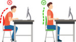
  • Sit up straight – Proper posture is important. Your back should be straight and your feet on the floor while you work. Elevate your wrists slightly instead of resting them on the keyboard.


  • Proper lighting – A setting that’s too bright (sunny backyard) or too dim (cavernous basement) can cause eye strain and headaches. Your screen should be bright enough that you don’t need to squint. A screen shield can help reduce glare.


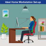
  • Set up monitor properly – Make sure your computer screen is about 25 inches, or an arm’s length, away from your face. The center of the screen should be about 10-15 degrees below eye level.  Cut glare by using a matte screen filter. You can find them for all types of computers, phones, and tablets.  Increase font size or set the magnification of the documents you are reading to a comfortable size.


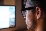
  • Consider computer glasses –For the greatest comfort at your computer, you might benefit from having your eye doctor modify your eyeglasses prescription to create customized computer glasses. This is especially true if you normally wear distance contact lenses, which may also become dry and uncomfortable during extended screen time.  Computer glasses also are a good choice if you wear bifocals or progressive lenses, because these lenses generally are not optimal for the distance to your computer screen.
  • Create Technology-Free Zones – These tips can help reduce eye strain when you’re forced to look at screens all day, creating technology-free zones in certain areas of your home, like the bedroom or bathroom. If you spend the entire day working on the computer, getting in bed and scrolling through social media until you fall asleep won’t do your eyes any favors.

Once you’re done for the day, truly unplug. Read a book or spend some quality time with family members without your phone.


  • Get an Eye Exam – If you have tried all these tips and eye strain is still an issue, it might be time to see an eye care professional to schedule an eye exam.  The exam may even detect underlying issues before they becomes worse.


Low Vision Awareness Month

Low vision affects millions of Americans — including many older adults. People with low vision aren’t blind, but because of their vision loss, they may not be able to do everyday tasks like driving or reading even with glasses.

What is Low Vision?

When your eyesight is impaired to the degree that you struggle with daily tasks like reading or cooking – or that you have difficulty recognizing faces, you may have a medical condition referred to as low vision. Someone with low vision can’t simply put on a pair of glasses or contacts and see well; this condition is beyond the typical loss of vision that occurs with aging.

Individuals experiencing low vision can struggle with maintaining independence; hobbies, reading and even socializing may become challenging as vision loss progresses. Because of the impact low vision can have on your life, it is important to have your eyes checked by a low vision specialist if you have any concerns.

Since it is unusual to be able to restore vision once it is lost, screenings can help preserve the vision you have and help you access adaptive lenses and devices if you need them. While there is not a cure for low vision, your specialist can help you adapt and if needed, create a vision rehabilitation program designed to meet your specific needs.

Low vision is often caused by one of these conditions:

  • Age-related macular degeneration (AMD) –AMD is a progressive eye condition affecting as many as 15 million Americans. The disease attacks the macula of the eye, where our sharpest central vision occurs, affecting reading, driving, identifying faces, watching television, safely navigating stairs and performing other daily tasks. Although it rarely results in complete blindness, it robs the individual of all but the outermost, peripheral vision, leaving only dim images or black holes at the center of vision. Read More (link to DEF website)
  • Diabetes Diabetic retinopathy is the most common diabetic eye disease and a leading cause of blindness in American adults caused by changes in the blood vessels of the retina.

In some people with diabetic retinopathy, blood vessels may swell and leak fluid or blood inside the eye. In other people, abnormal new blood vessels grow on the surface of the retina. The retina is the light-sensitive tissue at the back of the eye. A healthy retina is necessary for good vision. Read More (link to DEF website)

  • Glaucoma glaucoma is a disease that causes damage to the major nerve of the eye called the optic nerve, a part of the central nervous system that carries visual information from the eye to the brain.

The eye experiences a gradual increase of intraocular pressure (IOP) due to an imbalance of the fluid produced in the eye and the amount of fluid drained. Over time, elevated IOP can cause vision loss. The most common form of glaucoma is primary open angle glaucoma which affects about 3 million Americans. However, there are other types including narrow angle, congenital, normal tension, and secondary glaucoma. (link to DEF website)

  • Injury to the eye can also be a cause

Regular eye exams are essential for all adults; it is recommended that adults over the age of 60 have eye exams each year. For older adults, diabetics and anyone with the risk factors above, a visit with a low vision specialist may be warranted as well. This specialist differs from a conventional optometrist and is trained to detect low vision and offer solutions to help you retain or regain independence and the ability to do things you enjoy again.

While Low Vision Awareness Month is the ideal time to learn about this condition and about the risk factors you could be facing, you should get in touch with a low vision specialist any time you have concern about your eyesight. Any time you notice a change in vision, if you receive a new diagnosis or have one or more of the risk factors detailed above you should schedule an appointment right away.

Low Vision Resources from the National Eye Institute

It can be hard to describe low vision. Visit our website Vision Simulation to see what life is like with common causes of low vision, like age-related macular degeneration, diabetic retinopathy, and glaucoma.

Thanksgiving Dinner for your EYES!

Thanksgiving is almost here; a meal that nourishes the family bonds and traditions.  It’s the one time of the year where you can guarantee your eyes will be bigger than your stomach. This meal also has another added bonus — almost every item on the Thanksgiving table is healthy for your eyesight!

Here are several of the most popular Thanksgiving dishes and their corresponding benefits to your eye health:

Turkey –

No Thanksgiving is complete without a turkey, roasted golden brown and stuffed with fresh vegetables and herbs. Turkey is loaded with zinc and B-vitamin niacin, which helps prevent the formation of cataracts. Cataracts are the leading cause of vision loss in the United States, so gobble up!  You are “preventing” cataracts with every bite.


Spinach, Green Bean Casserole, Asparagus and Brussel Sprouts –

These foods and other leafy greens are loaded with lutein and zeaxanthin, two nutrients that protect the retina, which may also help reduce the risk of cataracts and macular degenerationHealthy Green Bean Casserole Recipe


Sweet Potatoes –

Sweet potatoes are full of Beta-carotene, which is a carotenoid and antioxidant that promotes night vision and overall good eyesight. Sweet potatoes are also loaded with vitamins C and E. Diets that are rich in these vitamins can help prevent or delay the development of cataracts and macular degeneration. Mashed Sweet Potatoes Recipe


Cranberry Sauce –

Cranberries contain bioflavonoids, a large class of antioxidants. Bioflavonoids are found in the pulp, skin and rinds of foods that contain vitamin C. Both flavonoids and vitamin C help protect the eyes from free radical damage caused by pollution and the body’s normal metabolic processes.


Pumpkin Pie –

Pumpkin is one of the best sources of vitamin A, so pumpkin pie is an eye-healthy dessert. In fact, one cup of cooked, mashed pumpkin contains more than 200 percent of the recommended daily intake of Vitamin A. Vitamin A provides nourishment and protection to the eye’s lens, cornea and macula (part of the retina), so it improves night vision. 


A diet that’s full of the right nutrients is a great start to keeping your eyes healthy, but don’t forget that regular eye exams are equally important! We hope you enjoy a happy and delicious Thanksgiving with family and friends.

*For more eye healthy recipes click here EYE COOK

Blinking – Why is it so important?

Blinking is something we all do every minute, whether we think about it or not. The average person blinks 15-20 times per minute, or about 1,200 times per hour.

So why do we unconsciously move our eyelids over our eyeballs so many times? Because it’s extremely important for your eyes and vision.

Although you may not think of blinking as a major component of your health care routine, if you didn’t blink for extended periods you’d be at higher risk of eye infection.  You would also have uncomfortable, dry eyes and would have decreased clarity of vision. The most important benefit of blinking is that it cleans the surface of your eye of any debris and washes it away with fresh tears. The thin coating of tears helps sharpen your vision, clearing and brightening the image your retina receives. Blinking also nourishes your eye with oxygen and nutrients, keeping your eyes healthy and comfortable.

Blinking wipes and renews your tear film, the smooth, moist layer covering our eyeballs.

The tears have 3 important layers.  The inner most is the mucous layer that contains ‘mucins’ to keep the tears attached to the surface cells of the cornea.  This material is produced by ‘goblet cells’ in the conjunctiva.  The next layer is aqueous or ‘water-like’ that is produced by the lacrimal gland located in the upper-outer quadrant of the eye region. Finally, the outer layer is lipid or ‘oil-like’ that prevents the other layers from evaporating. These lipids are produced by tiny glands along the lid margins, next to the eyelashes.  If any of these layers are absent or abnormal, then the cornea becomes vulnerable to damage.

The tears contain many protein components such as lysozyme, which function like natural antibiotics. The tear film also contains many nutrients and amino acids to nourish the cells of the cornea, which is the clear, dome-like structure on the front of the eye.

Besides keeping your eyes lubricated, the tear film also:

  • Helps form an almost perfectly smooth front optical surface on the cornea helping light to focus properly.
  • Washes away debris.
  • Transfers oxygen from the atmosphere to the cornea, since the cornea lacks blood vessels to deliver it directly.
  • Provides a pathway for white blood cells when there is an injury to the eye’s surface.
  • Prevents infection due to presence of lysozyme and other antibacterial enzymes.

Your eyelids also play a significant role, aside from protecting your eye and keeping things dark while you sleep. There is also a slight horizontal movement of the eyelid that pushes debris toward the tear ducts, which are the small openings at the inner corners of the eyes through which tears drain into your nose. That is why your nose runs when you cry.

Blinking exercises are a great way to keep your eyes moist. Blinking stimulates tear production and gives our eyes a chance to wash away any debris that has accumulated on the surface of the eye. Individuals who spend a lot of time on the computer or reading a lot tend to blink less often than they should. Therefore, if your eyes feel dry and irritated when using the computer or reading, then look away and do the following blinking exercises.

Blinking Exercises for Dry and Irritated Eyes:

  1. Close your eyes quickly and tightly. Squeeze your facial muscles around your eyes for three to five seconds. Open your eyes slowly and relax your facial muscles. Keep your eyes open for another three to five seconds and repeat the squeezing process five to seven times.
  2. Use the 10-10 rule and every 10 minutes look at away from your computer and intentionally blink 10 times. Then resume your activities.

WARZONE

A SPECIAL MESSAGE FROM DEF’S AMBASSADOR OF VISION

At this writing, the death toll from CoronaVirus (COVID-19) has just surpassed 60,000 with over 1 million cases reported in the US. It is almost impossible to comprehend the scope of this horrific pandemic, and the price that is being paid by healthcare professionals along with so many families who are unable to say goodbye to their loved ones who have often been forced to die alone.

At the same time, it was striking to learn that the four month death toll from the virus has just eclipsed the number of lives lost in the entire Vietnam War. We have heard the pandemic described as a war fought against a hidden enemy. While healthcare professionals confront the enemy in overcrowded hospitals across our country, there are others working just as hard in search of solutions that will allow us to return to our treasured normal way of life.

Researchers are quiet heroes laboring in laboratories in virtual anonymity, but with the same urgency as the healthcare professionals serving on the front lines of the battlefield. Whether it is in search of a vaccine or of a drug that may increase the possibility of overcoming the virus, the Discovery Eye Foundation is presently funding some of the critical efforts. As in past wars, all of us must do our part in the effort to defeat an enemy that continues its death march across the entire American landscape. No city or town, state or county, is immune from the icy hand of this most Grim Reaper.

I was attending college at the height of the Vietnam War, and I remember watching the evening news as the death toll continued to mount. Somehow, I found myself numb and somewhat insulated even as the numbers of casualties kept rising. It all changed when two of my friends from high school were lost during the 1968 Tet Offensive. Death became oh-so-real. We cannot allow the constant media exposure to dampen our awareness and our commitment to finding a cure and achieving complete victory in this all-out war.

When you contribute to Discovery Eye Foundations COVID-19 Emergency Fund, your funds go directly into the hands of the researchers. Even more important, their work will begin immediately. Your generosity has allowed us to be difference makers in the preservation of vision. Please join us as we enter the fight and turn our attention to finding answers to this most lethal and present danger.

To help the Discovery Eye Foundation join the fight against COVID-19, donate today!

DonateNow

Tom Sullivan
DEF’s Ambassador of Vision

sullivanvision.com

OUR EYES WORK LIKE CAMERA’S!

The inner workings of the human eye are complex, but at the same time, fascinating. Have you wondered how exactly they do work or what are the major parts of the eyeball involved in creating vision? Let’s find out.

These tiny cameras spend every day processing millions of pieces of information at lightning fast speeds, and turn them into the simple images we see almost instantly.

In reality, this process is anything but simple. The eye has several distinct parts, each of which has specific responsibilities that work together like a machine.

The eyeball is just like a camera. In fact, human eyes are part of a classification known as “camera-type eyes.” And just like a camera, it can’t function without the presence of light.  As light hits the eyes, it’s focused by the eye in a way similar to a camera lens. This process allows the images we see to appear clear and sharp rather than blurry.

There are specific parts of the eye that make this focusing process possible. Each beam of light that hits the eye goes through a series of steps:

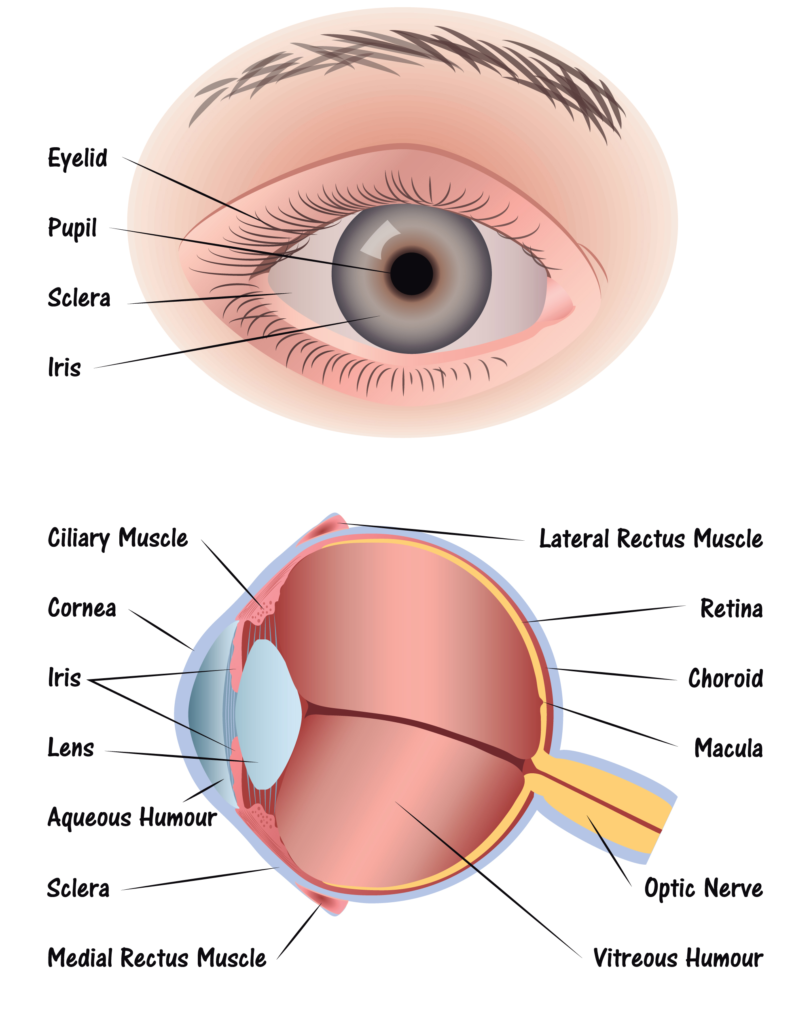
Step 1: Light passes through a thin layer of moisture

Step 2: Light hits the cornea.  The cornea is transparent, and is the first layer to begin focusing light within the eye. The cornea is connected to the sclera, which is a tough fiber on the outside of the eye that acts as protection.

Step 3: Behind the cornea is another liquid layer known as the aqueous humor, and its job is to maintain pressure levels in the front of the eye as light is passing through.

Step 4: Once light has passed through the aqueous humor, it has finally reached the pupil. The pupil is the round entryway of the colored iris.

Step 5: Once the pupil determines how much light it will let inside your eye, the job passes to the lens. The lens factors in the amount of light the pupil lets in, and figures out how far away you are from the object that the light is reflecting off of, or the object you’re trying to see. From there, the lens focuses your image into an accurate view of what you’re looking at. Part of this process is controlled by muscles in the lens called ciliary muscles, which expand and contract to pull on the lens and allow it to focus properly.

Step 6: As light reaches the center of the eye passes through another layer of moisture, called the vitreous, or vitreous humor. Then, it reaches the final stop in the process: The Retina.

The retina is the back of the eye. If the lens in your eye is most like a camera, the retina is most like its film – this is where the final product is projected. The retina has several parts:

  • Macula: The center of the retina. The center point of the macula is called the fovea, and it has the most photoreceptors and nerve endings of any part of the eye.
  • Photoreceptors: Split into two designations – rods and cones.
    • Cones are in the macula. When there is bright light, cones provide clear, sharp central vision and detect colors and fine details.
    • Rods are located outside the macula and extend all the way to the outer edge of the retina. They provide peripheral or side vision. Rods also allow the eyes to detect motion and help us see in dim light and at night.

  • Retinal pigment epithelium: Abbreviated RPE, this is a tissue layer below the rods and cones which absorbs any extra, unneeded light.
  • Choroid: The choroid is behind the retina, and is in charge of making sure the retina and RPE have enough nutrition flowing from small blood vessels.

Once the photoreceptors have converted light into an electronic signal, they send a signal to the brain’s visual command center and you have vision. It’s amazing what even small parts of our bodies can do.


How the Eye Works

January is Glaucoma Awareness Month

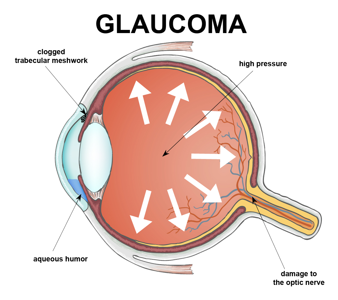
National Glaucoma Awareness Month reminds all of us to get regular eye exams and show support for those suffering from this conditionGlaucoma is one of the leading causes of vision loss and blindness.  Glaucoma is often called “the sneak thief of sight” because glaucoma has few symptoms or warning signs in its early stages. It can be diagnosed only by a full eye exam by an eye care practitioner.

Glaucoma is a disease where pressure builds up and damages the eye’s optic nerve. Types of this disease include the common Primary Open Angle Glaucoma, which causes peripheral eyesight to slowly diminish and is age-related. Angle Closure Glaucoma, where the fluid drainage system is narrow and closed so that the aqueous fluid remains in the front chamber of the eye and intraocular pressure rises; and Low Tension Glaucoma, where the optic nerve becomes damaged in spite of the intraocular pressures being within the normal range. There’s currently no way to restore vision lost from glaucoma because once the nerve cells become damaged, they do not regenerate.


A few important facts you should know about Glaucoma in adults:

  • More patients than ever are affected – Over 3 million people in the U.S. have glaucoma, and the number is rising.
  • Glaucoma can affect people of all ages – The most common form of glaucoma, Primary Open Angle Glaucoma, becomes more prevalent with increasing age. However, glaucoma can strike anyone, even infants and children but it is rare.
  • Demographics do play a role – Glaucoma is a leading cause of blindness among African Americans; it’s also highly prevalent in Hispanics over the age 65.
  • Is glaucoma hereditary? The risk of developing Primary Open–Angle Glaucoma is up to nine times more likely if parents or siblings have the disease.
  • Hope for future glaucoma patients – Although there is no cure for any form of glaucoma, early diagnosis and treatment help control the disease and slow the process of vision loss or blindness.

Newer Glaucoma Treatments 

Glaucoma treatment usually begins with the use of topical (eye drop) medications which lower the intraocular pressure. Within the past two years, two new topical medications have been approved for the treatment of glaucoma: VYZULTA® and Rhopressa®. VYZULTA® is a modification of a class of medications currently used to treat glaucoma – the prostaglandin analogs. This drug helps lower intraocular pressure by increasing the drainage of fluid (aqueous humor) from the eye.

Rhopressa® is part of a new class of drugs used to treat glaucoma called Rho kinase inhibitors. Rhopressa®, like VYZULTA®, also lowers intraocular pressure by increasing aqueous outflow. Both Rhopressa® and VYZULTA® are dosed once daily and pose few, if any, systemic safety concerns.
 

Laser

Using a laser to make a small opening in the iris to help with fluid drainage usually cures Angle-Closure Glaucoma.  This procedure is called a laser peripheral iridotomy.

For Primary Open Angel Glaucoma, when eye drops are not enough to reduce the pressure then a procedure called Selective Laser Trabeculoplasty (SLT) can be used. The SLT reduces intraocular pressure by stimulating increased outflow of fluid from the eye. SLT offers an improved safety profile compared to older glaucoma laser therapies and may lower eye pressure by as much as 20 to 30 percent. It is typically used as the next step in patients whose glaucoma is uncontrolled on medical therapy. Because of its excellent benefit-to-risk profile, however, SLT can sometimes be used in place of medications, especially in patients who have difficulty with their eye drops.
 

Surgery

When other treatments fail, there are many surgical therapies to lower the eye pressure. These surgical approaches, which are riskier than medical therapy or lasers, are usually employed when non-surgical means do not work well enough to stop vision loss.
 

MIGS

The goal of Minimally Invasive Glaucoma Surgery (MIGS) is to reduce intraocular pressure by enhancing the eye’s own internal aqueous humor drainage system. Some MIGS can be performed as stand-alone procedures, while others are typically done along with cataract surgery in patients with visually significant cataracts and mild to moderate glaucoma.

There are now a variety of recently FDA approved MIGS available for use in this country. MIGS typically are performed through a small incision in the eye with minimal tissue trauma and offer a favorable safety profile as well as more rapid visual recovery than traditional glaucoma surgery. There are many well qualified glaucoma specialists that perform the MIGS procedure.


Glaucoma Treatment Overview


National Glaucoma Awareness Month reminds all of us to get regular eye exams. Don’t let glaucoma steal your sight!  The best way to protect your sight is to get an annual comprehensive eye examination.


Thanks to funding from private philanthropists, DEF’s research continues to make great strides toward cures and treatment for glaucoma.  If you would like to support DEF’s sight saving research please donate today!

 DonateNow

Common Eye Problems in Winter

Winter time is here!! And as most people would expect, the holidays are a very popular time of the year. But the winter also comes with extreme temperatures, humidity and precipitation, so don’t let it affect your vision!

Be on the lookout for these common eye conditions this winter and hopefully, you will comfortably enjoy the season without any problems.


DRYNESS

Cold outdoor air and heated indoor air often have less moisture than other seasonal environments. In the winter, you may experience dry skin, chapped lips, and dry eyes due to this low humidity. Cold winter winds may also dry your eyes out. To learn more about dry eyes, visit the linked website at Dry Eyes.

To minimize the drying ability of winter air, keep yourself hydrated. Use non-preserved artificial tears several times a day. Running an humidifier in your home to improve the quality of your indoor air will help as well.


EXCESS TEARING

While some people experience a lack of tears in the winter, others have the opposite problem. Excess tearing and runny eyes can occur due to cold air, biting winds, or seasonal allergies. Pay attention to when your eyes tear up to determine the cause. If your eyes start to water when you step outside or when the wind blows your way, wear sunglasses or goggles to protect your eyes. Although it seems strange using additional artificial tears for teary eyes, nonetheless they can lessen this reflex tearing.

If you experience excess tearing and itching while indoors, try an allergy medication and appropriate eye drops to reduce the effect of seasonal allergies. If you cannot determine the cause of your watery eyes or if over-the-counter treatments have no effect, especially if the wateriness alters your vision, see an eye doctor.


LIGHT SENSITIVITY

Winter skies can seem dark and gloomy, but snowfall and ice create many reflective surfaces that can dramatically increase the amount of light that reaches your eyes. If you have sensitive eyes, you may experience even more blinking, discomfort, tearing and other symptoms in bright winter light.

Some individuals develop new light sensitivity during winter due to a condition known as “snow blindness”.  Always protect your eyes with UV filtering glasses or ski goggles when going outdoors for long periods of time, including when walking, shoveling snow, or other winter activities.


REDNESS

Harsh winter conditions can cause redness, tenderness, and inflammation in the eye area. You may have swollen eyelids or redness over the normally-white part of the eye (the conjunctiva which covers the white sclera).

This redness could result from dry eye or seasonal allergies. Use non-preserved artificial tears every 2-3 hours.  To reduce the discomfort of inflamed eyes, apply a cool compress, such as a damp washcloth and take an over-the-counter oral pain medication.  If your symptoms persist, see an eye doctor to determine the cause of the irritation.


VISION CHANGES

While many winter eye health problems result from increased light or decreased moisture, you can also experience eye conditions caused by cold temperatures.

If you notice vision changes while out in the cold, move to a warm area as soon as possible. Use non-preserved artificial tears every 2-3 hours to to keep your eyes moist. If your normal vision doesn’t return after 30 minutes or so, seek medical attention.


If you experience any of the seasonal problems listed above for a prolonged period, consult your eye doctor.